

Confirmation of long-term in vivo bearing mobility in eight rotating-platform TKAs
- PMID: 24771261
- PMCID: PMC4117897
- DOI: 10.1007/s11999-014-3642-6
Confirmation of long-term in vivo bearing mobility in eight rotating-platform TKAs
Abstract
Background: Posterior-stabilized rotating-platform prostheses for TKAs were designed to improve contact mechanics at the femoral-polyethylene (PE) interface. Short-term followup studies have shown that the PE bearings rotate with respect to the tibia but might not necessarily track with the femur. It is important to know how kinematics in these designs change owing to long-term in vivo use.
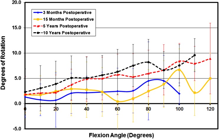
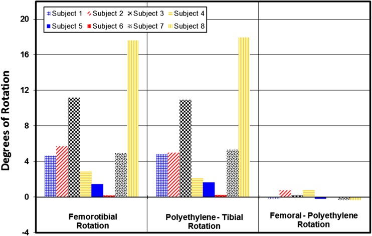
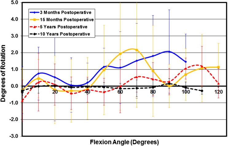
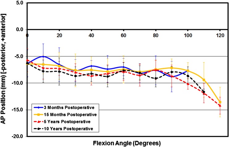
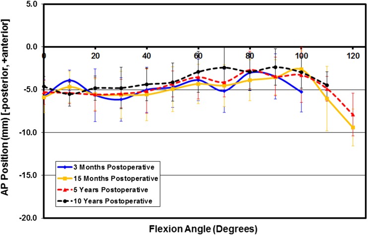
Questions/purposes: We asked whether there is a significant change in the in vivo kinematic performance of a posterior-stabilized rotating-platform prosthesis at as much as 10 years postoperative. We specifically examined (1) relative femoral component-PE bearing and relative PE bearing-tibial tray motion; (2) relative AP motion of the femoral condyles with respect to the tibial tray; and (3) relative femorotibial condylar translations.
Methods: In vivo three-dimensional kinematics were evaluated for eight patients at 3 months, 15 months, 5 years, and 10 years after TKA with primary implantation of a posterior-stabilized rotating-platform prosthesis. Each patient performed deep knee bend activity, and three-dimensional kinematics were reconstructed from multiple fluoroscopic images using a three-dimensional to two-dimensional registration technique. Once complete, relative component axial rotation patterns, medial and lateral condyle motions throughout flexion, and the presence of femoral condylar lift-off were analyzed.
Results: Overall, tibial bearing rotation was maintained at 10 years postoperatively. There was no statistical difference between postoperative periods for any kinematic parameter except for femoral component-PE bearing axial rotation, which was reduced at the 10-year evaluation versus other assessment periods (p = 0.0006). The lack of statistical difference between postoperative evaluation periods indicates sustained overall implant kinematic performance.
Conclusions: Our study showed that PE bearing-tibial tray mobility was maintained and that femoral component-PE bearing rotation was reduced at the 10-year followup. This suggests that the overall kinematic performance of this mobile-bearing implant is not negatively affected 10 years postoperatively.
Level of evidence: Level III, retrospective study. See the Instructions for Authors for a complete description of levels of evidence.
Figures
Similar articles
-
Mobile-bearing insert translational and rotational kinematics in a PCL-retaining total knee arthroplasty.Orthop Traumatol Surg Res. 2009 Jun;95(4):254-9. doi: 10.1016/j.otsr.2009.03.012. Epub 2009 May 12. Orthop Traumatol Surg Res. 2009. PMID: 19442597
-
In vivo kinematics comparison of fixed- and mobile-bearing total knee arthroplasty during deep knee bending motion.Knee Surg Sports Traumatol Arthrosc. 2014 Jul;22(7):1612-8. doi: 10.1007/s00167-012-2333-7. Epub 2012 Dec 12. Knee Surg Sports Traumatol Arthrosc. 2014. PMID: 23232786
-
Three-dimensional kinematics during deep-flexion kneeling in mobile-bearing total knee arthroplasty.Knee. 2011 Dec;18(6):412-6. doi: 10.1016/j.knee.2010.08.006. Epub 2010 Sep 15. Knee. 2011. PMID: 20833548
-
Rotating-platform TKA no different from fixed-bearing TKA regarding survivorship or performance: a meta-analysis.Clin Orthop Relat Res. 2014 Jul;472(7):2185-93. doi: 10.1007/s11999-014-3539-4. Epub 2014 Mar 4. Clin Orthop Relat Res. 2014. PMID: 24590838 Free PMC article. Review.
-
Influence of component design on in vivo tibiofemoral contact patterns during kneeling after total knee arthroplasty: a systematic review and meta-analysis.Knee Surg Sports Traumatol Arthrosc. 2021 Feb;29(2):446-466. doi: 10.1007/s00167-020-05949-y. Epub 2020 Apr 3. Knee Surg Sports Traumatol Arthrosc. 2021. PMID: 32242268
Cited by
-
Modifications of femoral component design in multi-radius total knee arthroplasty lead to higher lateral posterior femoro-tibial translation.Knee Surg Sports Traumatol Arthrosc. 2018 Jun;26(6):1645-1655. doi: 10.1007/s00167-017-4622-7. Epub 2017 Jun 27. Knee Surg Sports Traumatol Arthrosc. 2018. PMID: 28656456
-
The original Akagi line is the most reliable: a systematic review of landmarks for rotational alignment of the tibial component in TKA.Knee Surg Sports Traumatol Arthrosc. 2019 Apr;27(4):1018-1027. doi: 10.1007/s00167-018-5131-z. Epub 2018 Sep 10. Knee Surg Sports Traumatol Arthrosc. 2019. PMID: 30203197
-
Mobility of the rotating platform in low contact stress knee arthroplasty is durable.Knee Surg Sports Traumatol Arthrosc. 2017 Aug;25(8):2580-2585. doi: 10.1007/s00167-015-3823-1. Epub 2015 Oct 16. Knee Surg Sports Traumatol Arthrosc. 2017. PMID: 26475152
-
Are TKA Kinematics During Closed Kinetic Chain Exercises Associated with Patient-reported Outcomes? A Preliminary Analysis.Clin Orthop Relat Res. 2020 Feb;478(2):255-263. doi: 10.1097/CORR.0000000000000991. Clin Orthop Relat Res. 2020. PMID: 31634171 Free PMC article.
-
Posterior cruciate-retaining total knee arthroplasty exhibits small kinematic changes in the first postoperative year.Knee Surg Sports Traumatol Arthrosc. 2023 Mar;31(3):914-921. doi: 10.1007/s00167-022-07027-x. Epub 2022 Jun 16. Knee Surg Sports Traumatol Arthrosc. 2023. PMID: 35708746
References
MeSH terms
LinkOut - more resources
Full Text Sources
Other Literature Sources
Medical
Research Materials
