

Pathologic heterogeneity persists in early active multiple sclerosis lesions
- PMID: 24771535
- PMCID: PMC4070313
- DOI: 10.1002/ana.24163
Pathologic heterogeneity persists in early active multiple sclerosis lesions
Abstract
Objective: Multiple sclerosis (MS) lesions demonstrate immunopathological heterogeneity in patterns of demyelination. Previous cross-sectional studies reported immunopatterns of demyelination were identical among multiple active demyelinating lesions from the same individual, but differed between individuals, leading to the hypothesis of intraindividual pathological homogeneity and interindividual heterogeneity. Other groups suggested a time-dependent heterogeneity of lesions. The objective of our present study was to analyze tissue samples collected longitudinally to determine whether patterns of demyelination persist over time within a given patient.
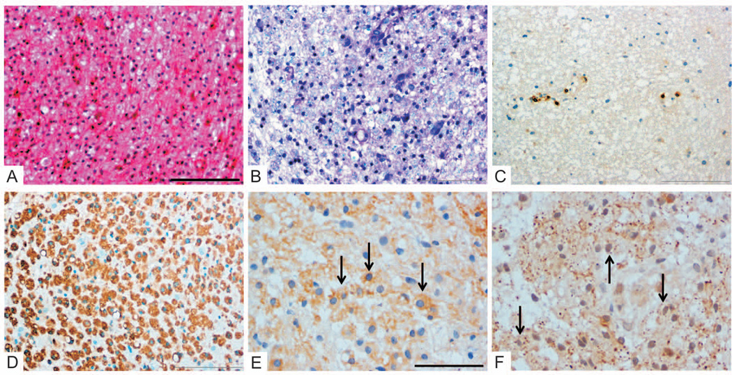
Methods: Archival tissue samples derived from patients with pathologically confirmed central nervous system inflammatory demyelinating disease who had undergone either diagnostic serial biopsy or biopsy followed by autopsy were analyzed immunohistochemically. The inclusion criteria consisted of the presence of early active demyelinating lesions--required for immunopattern classification--obtained from the same patient at 2 or more time points.
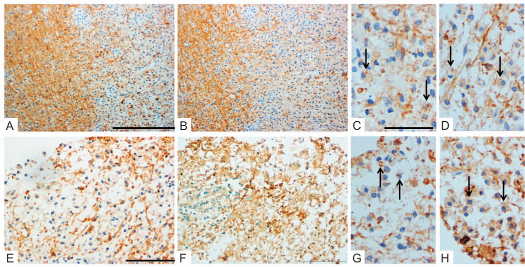
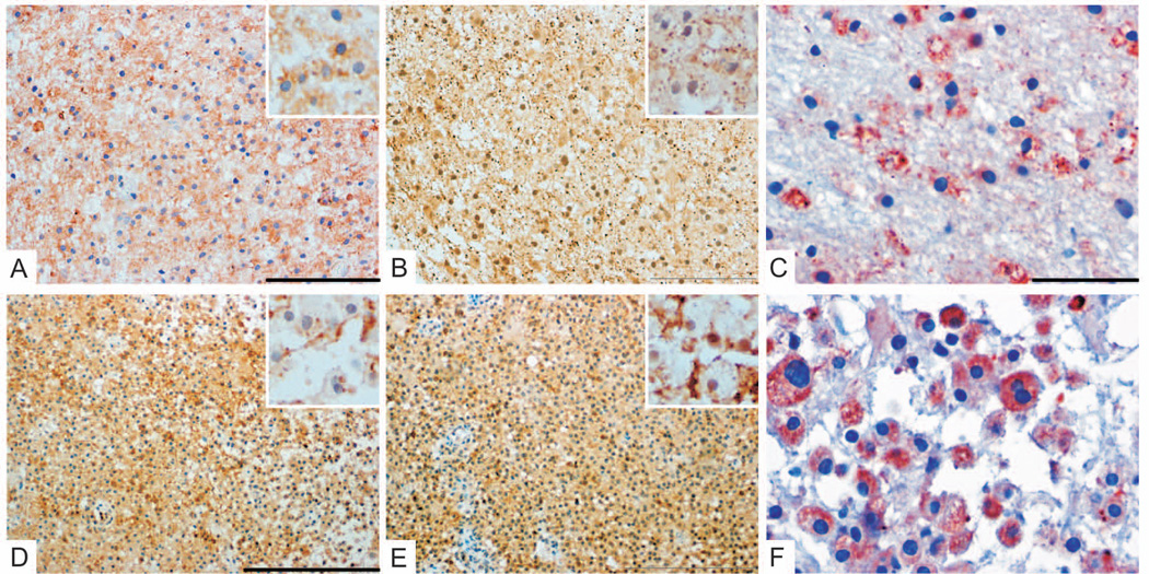
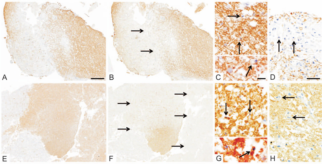
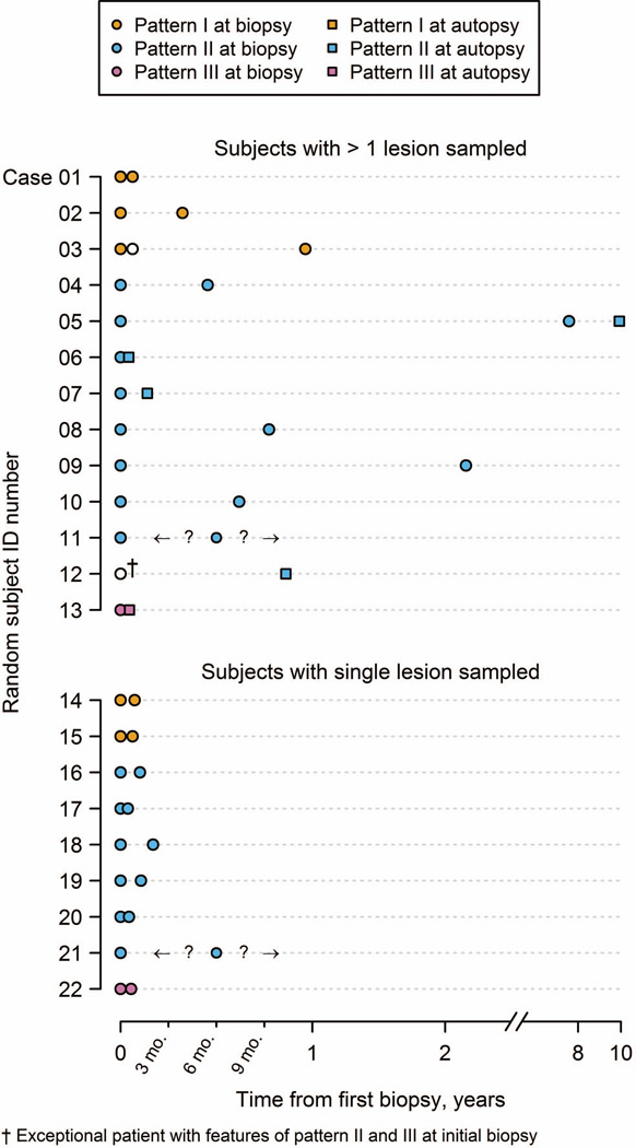
Results: Among 1,321 surgical biopsies consistent with MS, 22 cases met the study inclusion criteria. Twenty-one patients (95%) showed a persistence of immunopathological patterns in tissue sampled from different time points. This persistence was demonstrated for all major patterns of demyelination. A single patient showed features suggestive of both pattern II and pattern III on biopsy, but only pattern II among all active lesions examined at autopsy.
Interpretation: These findings continue to support the concept of patient-dependent immunopathological heterogeneity in early MS and suggest that the mechanisms and targets of tissue injury may differ among patient subgroups. These observations have potentially significant implications for individualized therapeutic approaches.
© 2014 American Neurological Association.
Conflict of interest statement
SDW, JMF, JEP, GY and HL have nothing to disclose.
Figures
References
-
- Anderson DW, Ellenberg JH, Leventhal CM, et al. Revised estimate of the prevalence of multiple sclerosis in the United States. Ann Neurol. 1992;31:333–336. - PubMed
-
- Brück W, Porada P, Poser S, et al. Monocyte/macrophage differentiation in early multiple sclerosis lesions. Ann Neurol. 1995;38:788–796. - PubMed
-
- Lucchinetti C, Brück W, Parisi J, et al. Heterogeneity of multiple sclerosis lesions: implications for the pathogenesis of demyelination. Ann Neurol. 2000;47:707–717. - PubMed
-
- Aboul-Enein F, Rauschka H, Kornek B, et al. Preferential loss of myelin-associated glycoprotein reflects hypoxia-like white matter damage in stroke and inflammatory brain diseases. J Neuropathol Exp Neurol. 2003;62:25–33. - PubMed
-
- Itoyama Y, Webster HD, Sternberger NH, et al. Distribution of papovavirus, myelin-associated glycoprotein, and myelin basic protein in progressive multifocal leukoencephalopathy lesions. Ann Neurol. 1982;11:396–407. - PubMed
Publication types
MeSH terms
Grants and funding
LinkOut - more resources
Full Text Sources
Other Literature Sources
Medical
Miscellaneous
