. 2014 Sep;115(9):1609-23.
doi: 10.1002/jcb.24824.

A protease storm cleaves a cell-cell adhesion molecule in cancer: multiple proteases converge to regulate PTPmu in glioma cells

Affiliations

A protease storm cleaves a cell-cell adhesion molecule in cancer: multiple proteases converge to regulate PTPmu in glioma cells

Polly J Phillips-Mason et al. J Cell Biochem. 2014 Sep.

Abstract

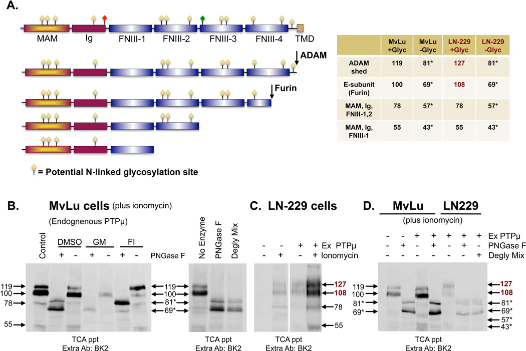
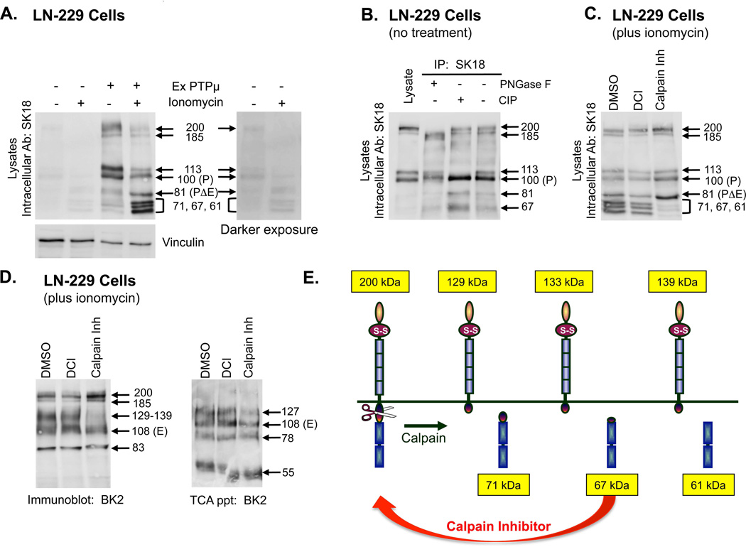
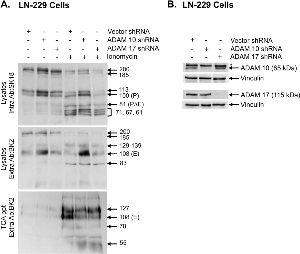
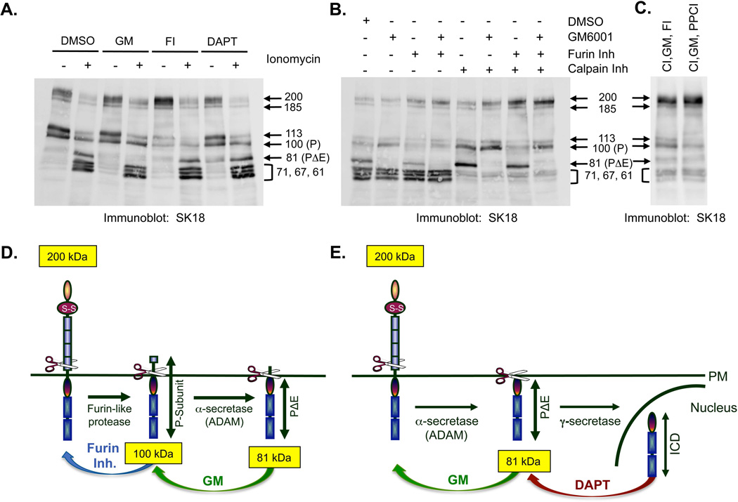
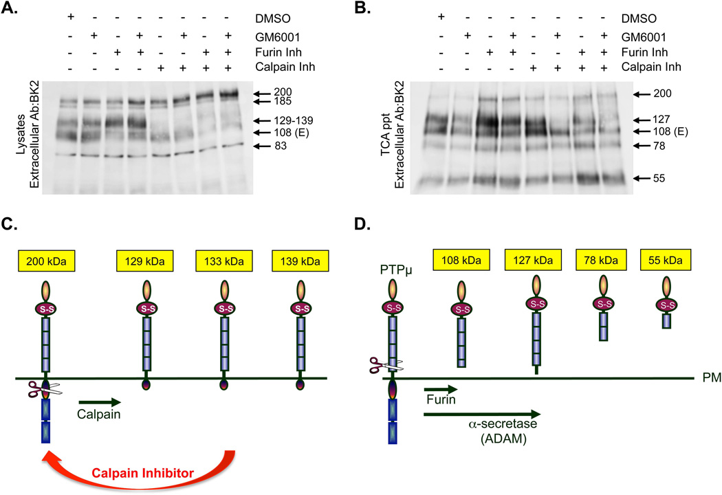
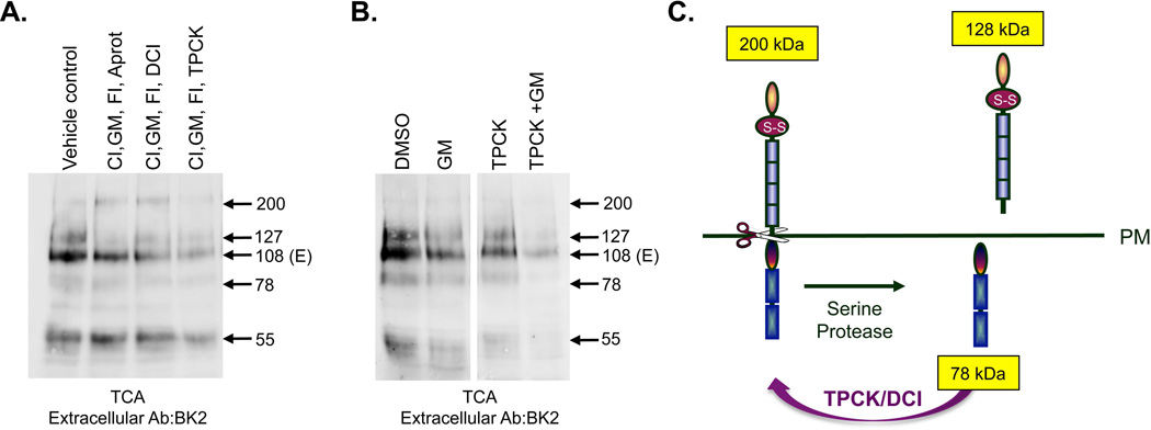
Cleavage of the cell-cell adhesion molecule, PTPµ, occurs in human glioblastoma multiforme brain tumor tissue and glioma cell lines. PTPµ cleavage is linked to increased cell motility and growth factor independent survival of glioma cells in vitro. Previously, PTPµ was shown to be cleaved by furin in the endoplasmic reticulum to generate membrane associated E- (extracellular) and P- (phosphatase) subunits, and by ADAMs and the gamma secretase complex at the plasma membrane. We also identified the presence of additional extracellular and intracellular PTPµ fragments in brain tumors. We set out to biochemically analyze PTPµ cleavage in cancer cells. We determined that, in addition to the furin-processed form of PTPµ, a pool of 200 kDa full-length PTPµ exists at the plasma membrane that is cleaved directly by ADAM to generate a larger shed form of the PTPµ extracellular segment. Notably, in glioma cells, full-length PTPµ is also subject to calpain cleavage, which generates novel PTPµ fragments not found in other immortalized cells. We also observed glycosylation and phosphorylation differences in the cancer cells. Our data suggest that an additional serine protease also contributes to PTPµ shedding in glioma cells. We hypothesize that a "protease storm" occurs in cancer cells whereby multiple proteases converge to reduce the presence of cell-cell adhesion molecules at the plasma membrane and to generate protein fragments with unique biological functions. As a consequence, the "protease storm" could promote the migration and invasion of tumor cells.

Keywords: PROTEOLYSIS; SHEDDING; PTPmu; RECEPTOR PROTEIN TYROSINE PHOSPHATASE; PROTEIN TYROSINE PHOSPHATASE; ADAM; CALPAIN; FURIN; SERINE PROTEASE; GLIOMA.


Figures

Figure 1
Figure 1. PTPμ is proteolytically processed by a Notch-like paradigm in Mv 1 Lu cells
Schematic of PTPμ proteolytic processing at the plasma membrane. Full-length (200 kDa) PTPμ is sequentially processed by a furin-like protease into two tightly associated fragments, the E-subunit (100 kDa) and the P-subunit (100 kDa). PTPμ is further processed by an α-secretase/ADAM to shed the E-subunit, resulting in a membrane tethered PΔE fragment (81 kDa) consisting of the entire intracellular segment and a few residues of the remaining extracellular segment. The PΔE fragment is a substrate for γ-secretase, which cleaves PTPμ within the transmembrane domain to release an intracellular fragment (ICD) capable of translocating to the nucleus (A). Mv 1 Lu cells were treated with ionomycin, after a 17–24 hour pre-incubation with DMSO, GM6001 or furin Inhibitor. Shed proteins were recovered from the culture supernatant by TCA precipitation and cell lyates were made as described in the Methods. Protein samples were resolved by SDS-PAGE and PTPμ was detected by immunoblotting with either the BK2 extracellular antibody or the SK18 intracellular antibody (B). Numbers indicate molecular weights in kDa. In this study we demonstrate that full-length PTPμ at the plasma membrane is a substrate for α-secretase/ADAM-induced ectodomain shedding in the absence of furin processing; producing a shed fragment of 119 kDa, and containing most of the extracellular segment (C).
Figure 2
Figure 2. PTPμ is differentially glycosylated in Mv 1 Lu and LN-229 cells
Extracellular fragments of PTPμ generated by furin and ADAM cleavage and the predicted PTPμ domains included in the 78 kDa and 55 kDa fragments, induced by ionomycin, are drawn based on size and antibody recognition. Meprin/PTPμ/A5 (MAM), immunoglobulin (Ig), fibronectin type III repeat (FNIII) and transmembrane (TMD) domains are indicated. Potential N-linked glycosylation sites based on NetNGlyc 1.0 are represented by yellow diamonds. Differential glycosylation was observed to “mark” the RPTP PTPkappa for proteolysis. Based on sequence homology, a similar site is present in PTPμ and is indicated by the red diamond. An O-linked glycosylation site predicted by NetOGlyc 1.0 exists in PTPμ as marked by a green diamond. The numbers indicate the observed sizes of the fragments when they are completely glycosylated or deglycosylated (asterisks). Numbers in burgundy represent the sizes of fragments observed in LN-229 cells that differ from those seen with Mv 1 Lu (A). Mv 1 Lu cells were treated with ionomycin, after a 17–24 hour pre-incubation with DMSO, GM6001 or furin inhibitor. Shed proteins were recovered from the culture supernatant by TCA precipitation. Protein samples were divided in half; one half was untreated, one half was subjected to deglycosylation by PNGase F. Proteins were resolved by SDS-PAGE and PTPμ fragments were detected using the BK2 antibody (B). TCA-precipitated shed proteins from Mv 1 Lu cell culture supernatant treated with ionomycin, was divided and either untreated, subjected to deglycosylation by PNGase F or to deglycosylation with a Deglycosylation Mix. Proteins were resolved by SDS-PAGE and PTPμ fragments were detected using the BK2 antibody (B, right panel). Parental LN-229 cells or LN-229 cells expressing exogenous PTPμ were either untreated or treated with ionomycin (C). Shed proteins were recovered from the culture supernatant by TCA precipitation and resolved by SDS-PAGE then detected by the BK2 antibody (C). Parental Mv 1 Lu cells, Mv 1 Lu cells expressing exogenous PTPμ and LN-229 cells expressing exogenous PTPμ were treated with ionomycin (D). Shed proteins were recovered from the culture supernatant by TCA precipitation and were subjected to deglycosylation by PNGase F or by a Deglycosylation Mix. Protein samples were resolved by SDS-PAGE and shed PTPμ detected by immunoblotting with BK2 (D). Numbers indicate molecular weights in kDa.
Figure 3
Figure 3. PTPμ is a substrate for calpain cleavage in LN-229 cells
Parental LN-229 cells or LN-229 cells expressing exogenous PTPμ were either untreated or treated with ionomycin for 30 minutes. Cellular protein lysates were made and proteins were resolved by SDS-PAGE as described in the Methods. Cell associated PTPμ was detected by SK18 (A). An antibody to vinculin was put on the SK18 immunoblot to show relative protein loading. A darker exposure of PTPμ expression in parental LN-229 cells plus and minus ionomycin is also shown (A). Protein lysates were made from untreated LN-229 cells expressing exogenous PTPμ and used for immunoprecipitations (IPs) with SK18. SK18 IPs were untreated or treated with CIP or PNGase F as described in the Methods and then resolved by SDS-PAGE. PTPμ full-length or intracellular fragments were detected with SK18 (B). LN-229 cells expressing exogenous PTPμ were treated with ionomycin after a 30 min pre-incubation with DMSO, DCI or calpain inhibitor. Cell-associated intracellular PTPμ was detected with SK18 in cellular protein lysates (C). Cell-associated extracellular fragments were detected with BK2 (D, left panel). Shed proteins were recovered from culture supernatant by TCA precipitation and resolved by SDS-PAGE and detected with BK2 (D, right panel). Numbers indicate molecular weights in kDa. A model of calpain cleavage of full-length PTPμ illustrates the observed products (E).
Figure 4
Figure 4. ADAM 10 regulates PTPμ shedding
Lentiviral shRNA constructs to ADAM 10, ADAM 17 and vector control were used to make lentiviral particles and infect LN-229 cells. Infected cells were either untreated or treated with ionomycin for 30 minutes. Cellular protein lysates were made and proteins were resolved by SDS-PAGE. Cell associated, full-length or intracellular fragments of PTPμ were detected in lysates with SK18 (top panel). Cell-associated extracellular fragments were detected in lysates by BK2 (middle panel). Shed proteins were recovered from the culture supernatant by TCA precipitation and resolved by SDS-PAGE were detected by the BK2 antibody (bottom panel) (A). Lysates from cells infected with lentiviral particles as described above, were resolved by SDS-PAGE and immublotted with polyclonal antibodies to either ADAM 10 or ADAM 17. The immunoblots were reprobed with a monoclonal antibody to vinculin to control for protein loading. The ADAM 10 iummunoblot shows a reduction in the 85 kDa precursor of ADAM 10 recognized by this antibody. The asteriks on the ADAM 10 immunoblot denotes a non-specific band. The ADAM 17 immunoblot shows a reduction in the 115 kDa band consistent with the expected molecular weight of ADAM 17 (B). Numbers indicate molecular weights in kDa.
Figure 5
Figure 5. Inhibition of proteolysis in LN-229 cells
Proposed PTPμ cleavage mechanisms in LN-229 cells with furin and ADAM (A) or calpain (B). LN-229 cells expressing exogenous PTPμ were either untreated or treated with ionomycin with or without a 17–24 hour pre-incubation with DMSO, GM6001 (25µM), furin inhibitor (30µM) or DAPT (1µM) (A). Cell-associated PTPμ in protein lysates resolved by SDS-PAGE was detected with SK18 (A). In B, LN-229 cells expressing exogenous PTPμ were treated with ionomycin after a 17–24 hour pre-incubation with DMSO, GM6001, furin Inhibitor or a 30 minute pre-incubation with calpain inhibitor (20µM), as single inhibitor treatments or in combination. Cell-associated PTPμ in protein lysates resolved by SDS-PAGE was detected with SK18 (B). LN-229 cells expressing exogenous PTPμ were treated with ionomycin after a pre-incubation with a combination of calpain inhibitor (CI), GM6001 (GM) and furin inhibitor (FI) or a combination of calpain inhibitor (CI), GM6001 (GM) and proprotein convertase inhibitor (PPCI, 25µM) (C). Cell-associated PTPμ in protein lysates resolved by SDS-PAGE was detected with SK18 (C). Numbers indicate molecular weights in kDa. PTPμ is a substrate for both furin and ADAM. The furin inhibitor stabilizes full-length PTPμ, and the ADAM inhibitor, GM6001, stabilizes the P-subunit (D). We determined full-length PTPμ is a direct substrate for ADAM cleavage. PΔE is generated in the presence of the furin inhibitor and is sensitive to the ADAM inhibitor, GM6001. PΔE is a substrate for constitutive cleavage by γ-secretase. Cleavage of PΔE by γ-secretase generates a labile ICD fragment of 78 kDa. The γ-secretase inhibitor, DAPT, can stabilize PΔE in the absence of ionomycin (E).
Figure 6
Figure 6. Multiple enzymes are involved in generating truncated membrane associated or ectodomain shed forms of PTPμ in LN-229 cells
LN-229 cells expressing exogenous PTPμ were treated with ionomycin after a 17–24 hour pre-incubation with DMSO, GM6001, furin inhibitor or a 30 minute pre-incubation with calpain inhibitor as single inhibitor treatments or in combination. Cellular protein lysates were made as described and resolved by SDS-PAGE. Cell-associated PTPμ was detected with the extracellular anitibody BK2 (A). Cells were treated as described above and the shed proteins were recovered from the culture supernatant by TCA precipitation. Shed proteins were resolved by SDS-PAGE and detected by the BK2 antibody (B). Numbers indicate molecular weights in kDa. Schematic of PTPμ membrane associated fragments generated by calpain cleavage induced by ionomycin (C). Calpain is predicted to cleave PTPμ at residues 825, 855 and 911. This diagram depicts the cell-associated extracellular fragments of PTPμ that would result from calpain cleavage and their predicted size (C). Schematic of the potential mechanisms used to generate the observed extracellular PTPμ fragments shed in response to ionomycin. PTPμ is a substrate for furin and ADAM. ADAM cleavage of furin-processed PTPμ generates a shed 100 kDa fragment. ADAM cleavage of full-length PTPμ generates a shed 127 kDa fragment in glioma cells. We also observe shed fragments of 78 and 55 kDa. These bands could arise either from full-length, furin-processed or ADAM shed PTPμ (D).
Figure 7
Figure 7. The combination of ADAM and serine protease inhibitors are required to block PTPμ cleavage
LN-229 cells expressing exogenous PTPμ were treated with ionomycin after a pre-incubation for 24 hours with GM6001 (GM), furin inhibitor (FI) combined with a 30 minutes pre-incubation with calpain inhibitor (CI) and Aprotinin, DCI, or TPCK. The vehicle control includes amounts of DMSO and methanol equal to the volume used for inhibitor delivery. Cell lysates were made and resolved by SDS-PAGE. Shed proteins were recovered from the culture supernatant by TCA precipitation, resolved by SDS-PAGE and detected by the BK2 antibody (A). Numbers indicate molecular weights in kDa. LN-229 cells expressing exogenous PTPμ were treated with ionomycin after a pre-incubation for 24 hours with DSMO or GM6001 (GM) combined with a 30 minute pre-incubation with TPCK. Cell lysates were analyzed for shed PTPμ as described above. (B). A model of serine protease cleavage of full-length PTPμ illustrates the observed products of a 128 kDa shed extracellular fragment and a 78 kDa intracellular fragment (C).

Similar articles

Cited by

References

    1. Anders L, Mertins P, Lammich S, Murgia M, Hartmann D, Saftig P, Haass C, Ullrich A. Furin-, ADAM 10-, and gamma-Secretase-Mediated Cleavage of a Receptor Tyrosine Phosphatase and Regulation of beta-Catenin's Transcriptional Activity. Mol Cell Biol. 2006;26:3917–3934. - PMC - PubMed
    1. Aricescu AR, Fulga TA, Cismasiu V, Goody RS, Szedlacsek SE. Intramolecular interactions in protein tyrosine phosphatase RPTPmu: kinetic evidence. Biochem Biophys Res Commun. 2001;280:319–327. - PubMed
    1. Brady-Kalnay SM, Flint AJ, Tonks NK. Homophilic binding of PTP mu, a receptor-type protein tyrosine phosphatase, can mediate cell-cell aggregation. J Cell Biol. 1993;122:961–972. - PMC - PubMed
    1. Brady-Kalnay SM, Mourton T, Nixon JP, Pietz GE, Kinch M, Chen H, Brackenbury R, Rimm DL, Del Vecchio RL, Tonks NK. Dynamic interaction of PTPmu with multiple cadherins in vivo. J Cell Biol. 1998;141:287–296. - PMC - PubMed
    1. Brady-Kalnay SM, Rimm DL, Tonks NK. Receptor protein tyrosine phosphatase PTPmu associates with cadherins and catenins in vivo. J Cell Biol. 1995;130:977–986. - PMC - PubMed

Publication types

MeSH terms