. 2014 Apr 22;2(4):e00283.
doi: 10.14814/phy2.283. Print 2014.

Mitochondrial mitophagy in mesenteric artery remodeling in hyperhomocysteinemia

Affiliations

Mitochondrial mitophagy in mesenteric artery remodeling in hyperhomocysteinemia

Anastasia Familtseva et al. Physiol Rep. .

Abstract

Abstract Although high levels of homocysteine also termed as hyperhomocysteinemia (HHcy) has been associated with inflammatory bowel disease and mesenteric artery occlusion, the mitochondrial mechanisms behind endothelial dysfunction that lead to mesenteric artery remodeling are largely unknown. We hypothesize that in HHcy there is increased mitochondrial fission due to altered Mfn-2/Drp-1 ratio, which leads to endothelial dysfunction and collagen deposition in the mesenteric artery inducing vascular remodeling. To test this hypothesis, we used four groups of mice: (i) WT (C57BL/6J); (ii) mice with HHcy (CBS+/-); (iii) oxidative stress resistant mice (C3H) and (iv) mice with HHcy and oxidative stress resistance (CBS+/-/C3H). For mitochondrial dynamics, we studied the expression of Mfn-2 which is a mitochondrial fusion protein and Drp-1 which is a mitochondrial fission protein by western blots, real-time PCR and immunohistochemistry. We also examined oxidative stress markers, endothelial cell, and gap junction proteins that play an important role in endothelial dysfunction. Our data showed increase in oxidative stress, mitochondrial fission (Drp-1), and collagen deposition in CBS+/- compared to WT and C3H mice. We also observed significant down regulation of Mfn-2 (mitochondrial fusion marker), CD31, eNOS and connexin 40 (gap junction protein) in CBS+/- mice as compared to WT and C3H mice. In conclusion, our data suggested that HHcy increased mitochondrial fission (i.e., decreased Mfn-2/Drp-1 ratio, causing mitophagy) that leads to endothelial cell damage and collagen deposition in the mesenteric artery. This is a novel report on the role of mitochondrial dynamics alteration defining mesenteric artery remodeling.

Keywords: Endothelial dysfunction; hyperhomocysteinemia; mitochondrial dynamics; oxidative stress.


Figures
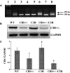
Figure 1.
Figure 1.
Genotypic and phenotypic analysis of WT, CBS+/− mice, C3H, and CBS+/−/C3H mice: (A) Lane 1: Molecular weight markers; Lane 2, 6&8: CBS+/− bands positioned at 450 and 308 bp; Lane 3, 4, 5&7: CBS+/+, band located at 308 bp; (B) Phenotype of WT, CBS+/−, C3H and CBS+/−/C3H mice with western blot using anti‐ CBS antibody, (C) Bar graphs for CBS protein expression, normalized with GAPDH, *P < 0.05 WT versus CBS+/−, #P < 0.05 CBS+/− versus C3H, §P < 0.05 WT versus CBS+/−/C3H, n = 4.
Figure 2.
Figure 2.
Oxidative stress status in mesenteric artery in HHcy: (A) Western blot analysis of Nox4, SOD‐1 and SOD‐2 protein expressions in WT, CBS+/−, C3H, CBS+/−/C3H mice mesentery. (B) Bar graph for respective protein in mesentery *P < 0.05 WT versus CBS+/−, #P < 0.05 CBS+/− versus C3H, §P < 0.05 CBS+/− versus CBS+/−/C3H, n = 4 per group (for two way ANOVA F > Fcrit., F = 25.71, P < 0.001). (C) Western blot analysis of eNOS protein expression in WT, CBS+/−, C3H, and CBS+/−/C3H mice mesentery. (D) Bar plot for eNOS protein expression normalized with GAPDH, *P < 0.05 WT versus CBS+/−, #P < 0.05 CBS+/− versus C3H, §P < 0.05 CBS+/− versus CBS+/−/C3H.
Figure 3.
Figure 3.
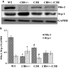
Mitochondrial dynamics in mesenteric artery in HHcy: (A) Western blot analysis of Mfn‐2 and Drp‐1 protein expressions in WT, CBS+/−, C3H, and CBS+/−/C3H mice mesentery. (B) Bar graph for Mfn‐2 and Drp‐1 protein expressions in mesentery, normalized with GAPDH, *P < 0.05 WT versus CBS+/−, #P < 0.05 CBS+/− versus C3H (for two way ANOVA F > Fcrit., F = 21.62, P < 0.0019).
Figure 4.
Figure 4.
Alteration of mitochondrial dynamics in mesenteric artery in HHcy: (A) Real‐time expression of Mfn‐2 mRNA in mesentery *P < 0.05 WT versus CBS+/−, #P < 0.05 CBS+/− versus C3H; (B) Real‐time expression of Drp‐1 mRNA in mesentery *P < 0.05 WT versus CBS+/−, #P < 0.05 CBS+/− versus C3H.
Figure 5.
Figure 5.
Increased mitochondrial fission in HHcy: (A) Mfn‐2 and Drp‐1 intensities in WT, CBS+/−, C3H, and CBS+/−/C3H mesenteric arteries with ×20 magnification. (B) Bar graph for Drp‐1 expression in mesenteric artery. Data expressed in arbitrary units. *P < 0.05 WT versus CBS+/−, #P < 0.05 WT versus CBS+/−C3H, §P < 0.05 C3H versus CBS+/−/C3H; for Mfn‐2 expression *P < 0.05 CBS+/− versus C3H.
Figure 6.
Figure 6.
Immunohistochemistry of mesenteric artery in different mouse groups: (A) CD31 and Connexin 40 intensities in WT, CBS+/−, C3H and CBS+/−/C3H mesenteric arteries, ×20 magnification. (B) Bar graph for respective proteins in mesenteric artery. Data expressed in arbitrary units. *P < 0.05 WT versus CBS+/−, #P < 0.05 CBS+/− versus C3H, §P < 0.05 CBS+/− versus CBS+/−/C3H, §§P < 0.05 WT versus CBS+/−/C3H, and C3H versus CBS+/−/C3H.
Figure 7.
Figure 7.
Masson Trichrome staining of mesenteric artery in different mouse strains: (A) Collagen intensity in mesenteric artery in WT, CBS+/−, C3H, and CBS+/−/C3H mice, ×20 magnification. (B) Bar graph for collagen expression in mesenteric arteries. Data expressed in arbitrary units. *P < 0.05 WT versus CBS+/−, #P < 0.05 CBS+/− versus C3H, §P < 0.05 CBS+/− versus CBS+/−/C3H.
Figure 8.
Figure 8.
Schematic representation of hypothesis: Hyperhomocysteinemia and oxidative stress alter mitochondrial dynamics by decreasing mitochondrial fusion and increasing fission, which leads to endothelial cell damage, mesenteric artery dysfunction, and vascular alterations with collagen accumulation.

References

    1. Chambers J. C., McGregor A., Jean‐Marie J., Obeid O. A., Kooner J. S. 1999. Demonstration of rapid onset vascular endothelial dysfunction after hyperhomocysteinemia: an effect reversible with vitamin C therapy. Circulation; 99:1156-1160 - PubMed
    1. Chen C., Conklin B. S., Ren Z., Zhong D. S. 2002. Homocysteine decreases endothelium‐dependent vasorelaxation in porcine arteries. J. Surg. Res.; 102:22-30 - PubMed
    1. Davidson S. M., Duchen M. R. 2007. Endothelial mitochondria: contributing to vascular function and disease. Circ. Res.; 100:1128-1141 - PubMed
    1. De Wit C., Roos F., Bolz S. S., Kirchhoff S., Kruger O., Willecke K. 2000. Impaired conduction of vasodilation along arterioles in connexin40‐deficient mice. Circ. Res.; 86:649-655 - PubMed
    1. Dranka B. P., Hill B. G., Darley‐Usmar V. M. 2010. Mitochondrial reserve capacity in endothelial cells: the impact of nitric oxide and reactive oxygen species. Free Radic. Biol. Med.; 48:905-914 - PMC - PubMed

LinkOut - more resources