. 2014 Jun 12;123(24):3760-9.
doi: 10.1182/blood-2013-08-521252. Epub 2014 Apr 25.

Runx1 exon 6-related alternative splicing isoforms differentially regulate hematopoiesis in mice

Affiliations

Runx1 exon 6-related alternative splicing isoforms differentially regulate hematopoiesis in mice

Yukiko Komeno et al. Blood. .

Abstract

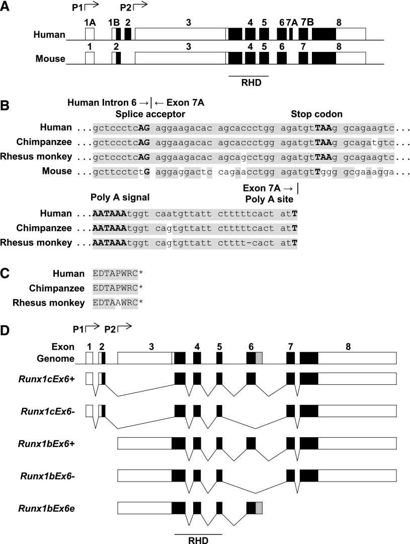
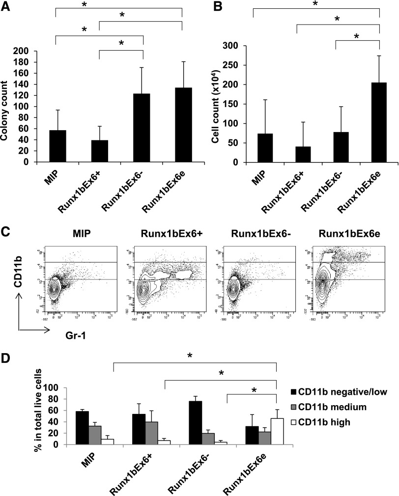
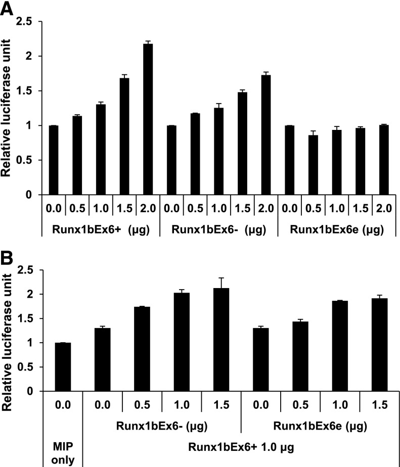
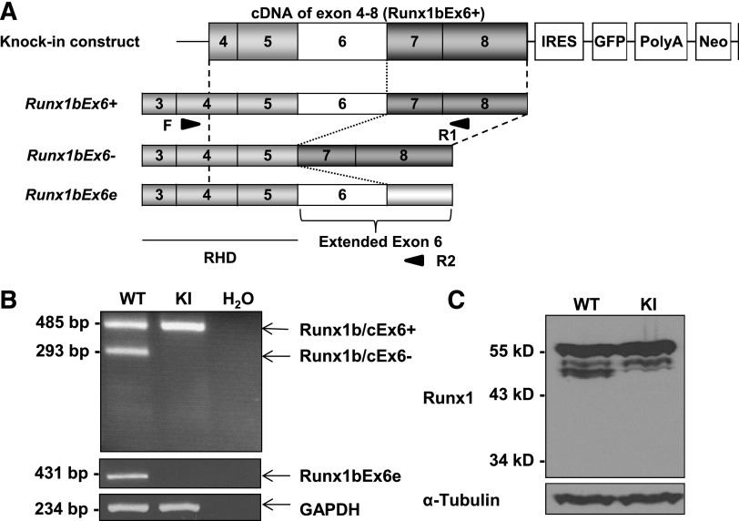
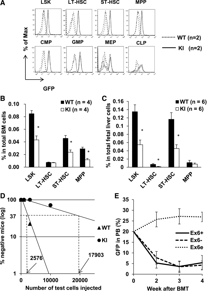
RUNX1 is an important transcription factor for hematopoiesis. There are multiple alternatively spliced isoforms of RUNX1. The best known isoforms are RUNX1a from use of exon 7A and RUNX1b and c from use of exon 7B. RUNX1a has unique functions due to its lack of C-terminal regions common to RUNX1b and c. Here, we report that the ortholog of human RUNX1a was only found in primates. Furthermore, we characterized 3 Runx1 isoforms generated by exon 6 alternative splicing. Runx1bEx6(-) (Runx1b without exon 6) and a unique mouse Runx1bEx6e showed higher colony-forming activity than the full-length Runx1b (Runx1bEx6(+)). They also facilitated the transactivation of Runx1bEx6(+). To gain insight into in vivo functions, we analyzed a knock-in (KI) mouse model that lacks isoforms Runx1b/cEx6(-) and Runx1bEx6e. KI mice had significantly fewer lineage-Sca1(+)c-Kit(+) cells, short-term hematopoietic stem cells (HSCs) and multipotent progenitors than controls. In vivo competitive repopulation assays demonstrated a sevenfold difference of functional HSCs between wild-type and KI mice. Together, our results show that Runx1 isoforms involving exon 6 support high self-renewal capacity in vitro, and their loss results in reduction of the HSC pool in vivo, which underscore the importance of fine-tuning RNA splicing in hematopoiesis.


Figures

Figure 1
Figure 1
Genome structure of Runx1 in various species. (A) Exon-intron structure of human RUNX1 and mouse Runx1. Genome sequences of Runx1 were collected from the GenBank database (NCBI). Boxes represent exons. Black boxes represent coding sequences. (B) Sequence homology of human RUNX1a intron 6 and exon 7A and their counterparts in other species. Top, Junction of human RUNX1a intron 6 and exon 7A. Bottom, Poly A signal and poly A site. Shaded areas show the common sequences. (C) Amino acid sequence encoded by human RUNX1a exon 7A and its counterparts in other species. Shaded area shows the common sequences. *, The end of the protein sequence. (D) Alternatively spliced isoforms of mouse Runx1. Boxes represent exons. Black boxes represent coding sequences. Gray box in Runx1bEx6e shows the part of extended exon 6 located in intron 6. P1, distal promoter; P2, proximal promoter.
Figure 2
Figure 2
Runx1bEx6 and Runx1bEx6e have higher colony-forming ability than full-length Runx1bEx6+. Lineage-negative BM cells were infected with indicated retroviral constructs. After 1-week selection in 1 μg/mL puromycin, 1 × 10⁵ cells were seeded into methylcellulose in duplicate. One week later, (A) colony counts and (B) cell counts per plate were evaluated. Mean ± SD of 5 independent experiments is shown. (C) Flow cytometric analysis of colony cells. The gating of CD11b-negative/low, -medium, and -high (from bottom to top) is shown in the plots. Representative result of 3 independent experiments is shown. (D) Quantification of the results in panel C. Mean ± SD of 3 independent experiments is shown.
Figure 3
Figure 3
Runx1bEx6 and Runx1bEx6e have lower transactivation efficiency than full-length Runx1bEx6+. 293T cells were transfected in duplicate with the indicated construct together with CBFβ expression construct, M-CSF receptor promoter-firefly luciferase reporter construct and Renilla luciferase reporter construct as a transfection efficiency control. (A) Transfection with single Runx1 isoform. Runx1bEx6 shows lower transactivation than Runx1bEx6+. Runx1bEx6e has no transactivation. Values were normalized to Renilla luciferase signal, and promoter activity of MIP-transfected cells was set to 1. Representative result of 2 independent experiments is shown. (B) Cotransfection of MIP, Runx1bEx6, or Runx1bEx6e with Runx1bEx6+. Runx1bEx6 and Runx1bEx6e show additive and synergistic effect, respectively. Values were normalized to Renilla signal, and MIP was set to 1. Representative result of 2 independent experiments is shown.
Figure 4
Figure 4
Runx1 isoforms have various protein stability and susceptibility to proteasome-mediated degradation. (A-B) Protein stability of Runx1 isoforms. 293T cells were transfected with indicated construct. Forty-eight hours later, cells were treated with 100 ng/mL CHX for indicated time. α-Tubulin served as a loading control. (A) Representative western blotting result of 3 independent experiments. (B) Quantification of the results in panel A. Intensities at 0 h were set to 100%. Mean ± SD of 2 independent experiments is shown. (C-D) Inhibition of proteasome-mediated degradation pathway. 293T cells were transfected with the indicated construct. Forty-eight hours later, cells were treated with 10 µM MG132 for 24 hours. α-Tubulin served as a loading control. (C) Representative western blotting result of 3 independent experiments is shown. (D) Quantification of the results in panel C. Runx1 signals were normalized to those of tubulin, and vehicle (DMSO)–treated signals were set to 1. Mean ± SD of 3 independent experiments is shown. (E) Western blot of phosphorylation of “S303”. Replicate membranes were probed with anti-Runx1 and anti-phospho S303 antibodies, respectively. (F) Quantification of panel E. Mean ± SD of 3 independent experiments is shown.
Figure 5
Figure 5
Runx1-IRES-GFP KI mice lack the expression of 3 Runx1 isoforms. (A) Structure of the Runx1-IRES-GFP KI construct. Boxes with numbers represent exons. Light gray boxes are RHD, and dark gray boxes are regulatory domains. Arrowheads show the positions of primers (F, R1, and R2) used for panel B. (B) Expression of Runx1 isoforms in total BM cells by RT-PCR. KI cells lack Runx1b/cEx6 and Runx1bEx6e. GAPDH serves as a loading control. Water lane is the negative control. Representative result of 3 independent experiments is shown. (C) Protein expression of Runx1 in lineage-depleted c-Kit–enriched BM cells. KI lane has the same level of the major band although it lacks smaller bands which correspond to Runx1b/cEx6. Runx1bEx6e band is undetectable in either WT or KI lane. α-Tubulin serves as a loading control. Representative result of 2 independent experiments is shown. Neo, neomycin resistance gene; PolyA, polyadenylation signal.
Figure 6
Figure 6
Runx1-IRES-GFP KI mice have decreased HSC pool. (A) GFP expression in HSPCs. GFP fluorescence in KI cells indicates Runx1 promoter activity. GFP histograms are shown by gating the indicated population. Dashed and solid lines denote WT and KI cells, respectively (2 mice each). (B) Flow cytometric analysis of adult BM. (C) Flow cytometric analysis of E14.5 fetal liver cells. (D) Competitive repopulation unit assay. The detailed protocol is described in supplemental Methods. WT cells have 7 times higher frequency of stem cells than KI cells. This experiment was repeated twice with similar results. (E) BMT of Runx1 overexpressing cells. One million cells with 20% GFP+ cells were injected into recipient mice. GFP percentage in peripheral blood was followed up. Ex6+, n = 4. Ex6, n = 3. Ex6e, n = 4. Ex6+, Ex6, and Ex6e represent Runx1bEx6+, Runx1bEx6, and Runx1bEx6e, respectively. LT-HSC, long-term HSC.

Comment in

References

    1. Miyoshi H, Shimizu K, Kozu T, Maseki N, Kaneko Y, Ohki M. t(8;21) breakpoints on chromosome 21 in acute myeloid leukemia are clustered within a limited region of a single gene, AML1. Proc Natl Acad Sci USA. 1991;88(23):10431–10434. - PMC - PubMed
    1. Lam K, Zhang DE. RUNX1 and RUNX1-ETO: roles in hematopoiesis and leukemogenesis. Front Biosci (Landmark Ed) 2012;17:1120–1139. - PMC - PubMed
    1. Zhang DE, Fujioka K, Hetherington CJ, et al. Identification of a region which directs the monocytic activity of the colony-stimulating factor 1 (macrophage colony-stimulating factor) receptor promoter and binds PEBP2/CBF (AML1). Mol Cell Biol. 1994;14(12):8085–8095. - PMC - PubMed
    1. Takahashi A, Satake M, Yamaguchi-Iwai Y, et al. Positive and negative regulation of granulocyte-macrophage colony-stimulating factor promoter activity by AML1-related transcription factor, PEBP2. Blood. 1995;86(2):607–616. - PubMed
    1. Huang G, Zhang P, Hirai H, et al. PU.1 is a major downstream target of AML1 (RUNX1) in adult mouse hematopoiesis. Nat Genet. 2008;40(1):51–60. - PubMed

Publication types