

Review
doi: 10.1016/j.cpet.2013.10.006.
Brain: normal variations and benign findings in fluorodeoxyglucose-PET/computed tomography imaging
- PMID: 24772054
- PMCID: PMC3998066
- DOI: 10.1016/j.cpet.2013.10.006
Item in Clipboard
Review
Brain: normal variations and benign findings in fluorodeoxyglucose-PET/computed tomography imaging
PET Clin.
2014 Apr.
Abstract
Brain 18F-fluorodeoxyglucose (18F-FDG) PET allows the in vivo study of cerebral glucose metabolism, reflecting neuronal and synaptic activity. 18F-FDG-PET has been extensively used to detect metabolic alterations in several neurologic diseases compared with normal aging. However, healthy subjects have variants of 18F-FDG distribution, especially as associated with aging. This article focuses on 18F-FDG-PET findings in so-called normal brain aging, and in particular on metabolic differences occurring with aging and as a function of people’s gender. The effect of different substances, medications, and therapy procedures are discussed, as well as common artifacts.
Figures
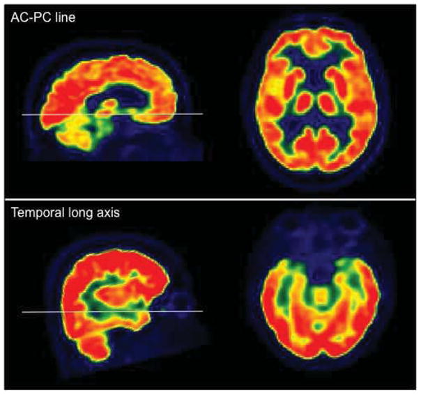
18F-FDG PET images oriented along AC-PC line and temporal long axis.
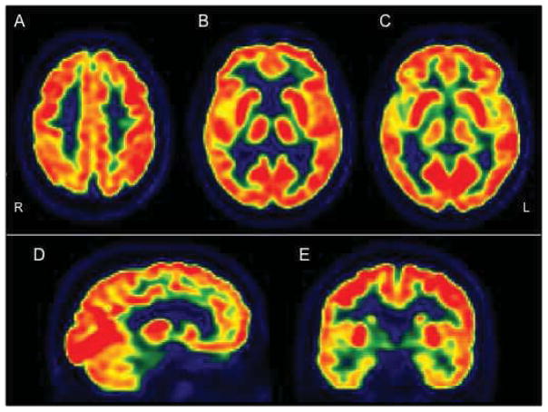
18F-FDG PET of a normal subject. A, B, C: transaxial plane, showing higher 18F-FDG uptake in basal ganglia, frontal eye fields, posterior cingulate and visual cortices. D: sagittal plane. E: coronal plane, showing lower 18F-FDG activity in temporal lobes, particularly in medial temporal cortex, as compared to the other regions.
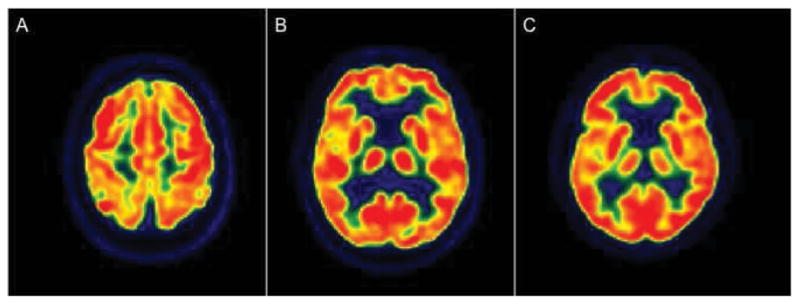
Age-related metabolic reductions in 3 cognitively normal subjects. A: 80 year-old subject, demonstrating prefrontal and superior parietal hypometabolism with sparing of primary sensory-motor cortices. B: 76 year-old subject, showing mild hypometabolism in anterior cingulate cortex, medial frontal regions and insula. C: 82 year-old subject, demonstrating hypometabolism involving anterior cingulate cortex, medial frontal regions and insula.
References
-
- Sokoloff L, Reivich M, Kennedy C, et al. THE [14C]DEOXYGLUCOSE METHOD FOR THE MEASUREMENT OF LOCAL CEREBRAL GLUCOSE UTILIZATION: THEORY, PROCEDURE, AND NORMAL VALUES IN THE CONSCIOUS AND ANESTHETIZED ALBINO RAT. Journal of Neurochemistry. 1977;28:897–916. - PubMed
-
- Sokoloff L. Energetics of functional activation in neural tissues. Neurochem Res. 1999;24(2):321–329. - PubMed
Publication types
MeSH terms
Substances
Grants and funding
LinkOut - more resources
Full Text Sources
Other Literature Sources
Medical