Review
. 2014 Apr 3:5:60.
doi: 10.3389/fphar.2014.00060. eCollection 2014.

Modeling CaMKII-mediated regulation of L-type Ca(2+) channels and ryanodine receptors in the heart

Affiliations
Review

Modeling CaMKII-mediated regulation of L-type Ca(2+) channels and ryanodine receptors in the heart

Joseph L Greenstein et al. Front Pharmacol. .

Abstract

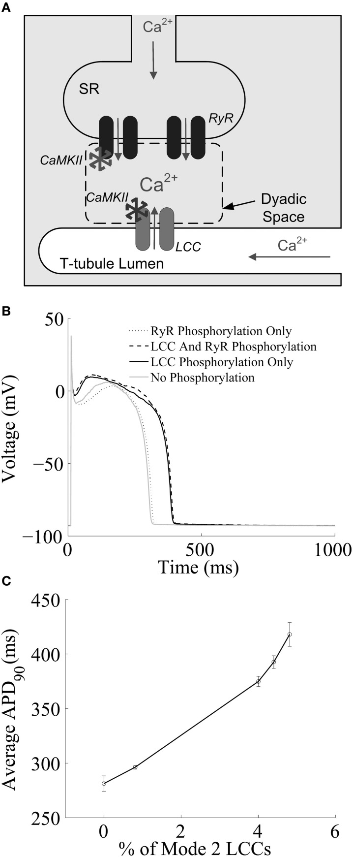
Excitation-contraction coupling (ECC) in the cardiac myocyte is mediated by a number of highly integrated mechanisms of intracellular Ca(2+) transport. Voltage- and Ca(2+)-dependent L-type Ca(2+) channels (LCCs) allow for Ca(2+) entry into the myocyte, which then binds to nearby ryanodine receptors (RyRs) and triggers Ca(2+) release from the sarcoplasmic reticulum in a process known as Ca(2+)-induced Ca(2+) release. The highly coordinated Ca(2+)-mediated interaction between LCCs and RyRs is further regulated by the cardiac isoform of the Ca(2+)/calmodulin-dependent protein kinase (CaMKII). Because CaMKII targets and modulates the function of many ECC proteins, elucidation of its role in ECC and integrative cellular function is challenging and much insight has been gained through the use of detailed computational models. Multiscale models that can both reconstruct the detailed nature of local signaling events within the cardiac dyad and predict their functional consequences at the level of the whole cell have played an important role in advancing our understanding of CaMKII function in ECC. Here, we review experimentally based models of CaMKII function with a focus on LCC and RyR regulation, and the mechanistic insights that have been gained through their application.

Keywords: CaMKII; cardiac myocyte; cell signaling; computational modeling; excitation-contraction coupling.


Figures

Figure 1
Figure 1
(A) Schematic representation of the cardiac dyad. Ca2+ ions pass through the LCC and enter the dyad, where they bind to RyRs, triggering CICR. The activity of each CaMKII monomer is a function of dyadic Ca2+ levels. Ca2+ ions released from the SR diffuse out of the dyad into the bulk myoplasm. (B) Simulated results from a 1-Hz AP-pacing protocol under different LCC and/or RyR phosphorylation conditions. (C) APD90 as a function of average LCC phosphorylation levels. Fully phosphorylated LCCs gate in mode 2, which exibit long-duration openings. (A) Reprinted with permission from Hashambhoy et al. (2009). Copyright 2009 Elsevier. (B,C) Reprinted with permission from Hashambhoy et al. (2010). Copyright 2010 Elsevier.
Figure 2
Figure 2
(A) State diagram of the stochastic CaMKII activation model of Foteinou et al. (2013). Prior to the introduction of Ca2+/CaM, all of the CaMKII subunits are in the inactive form (state I). Activation occurs upon binding of Ca2+/CaM (state B), autophosphorylation (state P), and oxidation (state OxB). Autonomous active states (Ca2+/CaM-unbound) can be either autophosphorylated (state A) or oxidized (state OxA). The model also includes an active state that is both oxidized and phosphorylated (state OxP). (B) Simulated steady state APs under control conditions (0 μM H2O2). (C) Simulated APs, some of which exhibit EADs, under conditions of elevated oxidant stress (200 μM H2O2). Simulated APs are from a 2 s PCL pacing protocol (ensemble of 12500 calcium release units). For each condition, results for 10 consecutive APs following an initial 10 s of pacing are shown.

References

    1. Anderson M. E., Braun A. P., Schulman H., Premack B. A. (1994). Multifunctional Ca2+/calmodulin-dependent protein kinase mediates Ca(2+)-induced enhancement of the L-type Ca2+ current in rabbit ventricular myocytes. Circ. Res. 75, 854–861 10.1161/01.RES.75.5.854 - DOI - PubMed
    1. Baddeley D., Jayasinghe I. D., Lam L., Rossberger S., Cannell M. B., Soeller C. (2009). Optical single-channel resolution imaging of the ryanodine receptor distribution in rat cardiac myocytes. Proc. Natl. Acad. Sci. U.S.A. 106, 22275–22280 10.1073/pnas.0908971106 - DOI - PMC - PubMed
    1. Bers D. M. (2001). Excitation-Contraction Coupling and Cardiac Contractile Force. Boston, MA: Kluwer; 10.1007/978-94-010-0658-3 - DOI
    1. Bhalla U. S., Iyengar R. (1999). Emergent properties of networks of biological signaling pathways. Science 283, 381–387 10.1126/science.283.5400.381 - DOI - PubMed
    1. Bondarenko V. E., Szigeti G. P., Bett G. C., Kim S. J., Rasmusson R. L. (2004). Computer model of action potential of mouse ventricular myocytes. Am. J. Physiol. Heart Circ. Physiol. 287, H1378–H1403 10.1152/ajpheart.00185.2003 - DOI - PubMed

LinkOut - more resources