Cerebrovascular and neuroprotective effects of adamantane derivative
- PMID: 24772429
- PMCID: PMC3977474
- DOI: 10.1155/2014/586501
Cerebrovascular and neuroprotective effects of adamantane derivative
Abstract
Objectives: The influence of 5-hydroxyadamantane-2-on was studied on the rats' brain blood flow and on morphological state of brain tissue under the condition of brain ischemia. The interaction of the substance with NMDA receptors was also studied.
Methods: Study has been implemented using the methods of local blood flow registration by laser flowmeter, [(3)H]-MK-801binding, and morphological examination of the brain tissue. We used the models of global transient ischemia of the brain, occlusion of middle cerebral artery, and hypergravity ischemia of the brain.
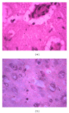
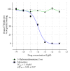
Results: Unlike memantine, antagonist of glutamatergic receptors, the 5-hydroxyadamantane-2-on does not block NMDA receptors but enhances the cerebral blood flow of rats with brain ischemia. This effect is eliminated by bicuculline. Under conditions of permanent occlusion of middle cerebral artery, 5-hydroxyadamantane-2-on has recovered compensatory regeneration in neural cells, axons, and glial cells, and the number of microcirculatory vessels was increased. 5-Hydroxyadamantane-2-on was increasing the survival rate of animals with hypergravity ischemia.
Conclusions: 5-Hydroxyadamantane-2-on, an adamantane derivative, which is not NMDA receptors antagonist, demonstrates significant cerebrovascular and neuroprotective activity in conditions of brain ischemia. Presumably, the GABA-ergic system of brain vessels is involved in mechanisms of cerebrovascular and neuroprotective activity of 5-hydroxyadamantane-2-on.
Figures


References
-
- Lo EH, Dalkara T, Moskowitz MA. Mechanisms, challenges and opportunities in stroke. Nature Reviews Neuroscience. 2003;4(5):399–415. - PubMed
-
- Lo EH, Moskowitz MA, Jacobs TP. Exciting, radical, suicidal: how brain cells die after stroke. Stroke. 2005;36(2):189–192. - PubMed
-
- Lok J, Gupta P, Guo S, et al. Cell-cell signaling in the neurovascular unit. Neurochemical Research. 2007;32(12):2032–2045. - PubMed
-
- Broughton BRS, Reutens DC, Sobey CG. Apoptotic mechanisms after cerebral ischemia. Stroke. 2009;40(5):e331–e339. - PubMed
-
- Galeffi F, Sinnar S, Schwartz-Bloom RD. Diazepam promotes ATP recovery and prevents cytochrome c release in hippocampal slices after in vitro ischemia. Journal of Neurochemistry. 2000;75(3):1242–1249. - PubMed
MeSH terms
Substances
LinkOut - more resources
Full Text Sources
Other Literature Sources

