Type III interferons are expressed by Coxsackievirus-infected human primary hepatocytes and regulate hepatocyte permissiveness to infection
- PMID: 24773058
- PMCID: PMC4137853
- DOI: 10.1111/cei.12368
Type III interferons are expressed by Coxsackievirus-infected human primary hepatocytes and regulate hepatocyte permissiveness to infection
Abstract
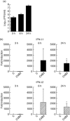
Hepatitis is a common and potentially fatal manifestation of severe Coxsackievirus infections, particularly in newborn children. Little is known of the immune-mediated mechanisms regulating permissiveness to liver infection. It is well established that type I interferons (IFNs) play an important role in the host innate immune response to Coxsackievirus infections. Recent studies have highlighted a role for another IFN family, the type III IFNs (also called IFN-λ), in anti-viral defence. Whether type III IFNs are produced by hepatocytes during a Coxsackievirus infection remains unknown. Moreover, whether or not type III IFNs protects hepatocytes from a Coxsackievirus infection has not been addressed. In this study, we show that primary human hepatocytes respond to a Coxsackievirus B3 (CVB3) infection by up-regulating the expression of type III IFNs. We also demonstrate that type III IFNs induce an anti-viral state in hepatocytes characterized by the up-regulated expression of IFN-stimulated genes, including IFN-stimulated gene (ISG15), 2'-5'-oligoadenylate synthetase 2 (OAS2), protein kinase regulated by dsRNA (PKR) and myxovirus resistance protein 1 (Mx1). Furthermore, our study reveals that type III IFNs attenuate CVB3 replication both in hepatocyte cell lines and primary human hepatocytes. Our studies suggest that human hepatocytes express type III IFNs in response to a Coxsackievirus infection and highlight a novel role for type III IFNs in regulating hepatocyte permissiveness to this clinically relevant type of virus.
Keywords: Coxsackievirus; anti-viral defence; enterovirus; hepatocytes; interferons.
© 2014 British Society for Immunology.
Figures


Similar articles
-
Interferon-beta expression and type I interferon receptor signaling of hepatocytes prevent hepatic necrosis and virus dissemination in Coxsackievirus B3-infected mice.PLoS Pathog. 2018 Aug 3;14(8):e1007235. doi: 10.1371/journal.ppat.1007235. eCollection 2018 Aug. PLoS Pathog. 2018. PMID: 30075026 Free PMC article.
-
Induction of an antiviral state and attenuated coxsackievirus replication in type III interferon-treated primary human pancreatic islets.J Virol. 2013 Jul;87(13):7646-54. doi: 10.1128/JVI.03431-12. Epub 2013 May 1. J Virol. 2013. PMID: 23637411 Free PMC article.
-
Over-expression of mitochondrial antiviral signaling protein inhibits coxsackievirus B3 infection by enhancing type-I interferons production.Virol J. 2012 Dec 19;9:312. doi: 10.1186/1743-422X-9-312. Virol J. 2012. PMID: 23249700 Free PMC article.
-
A Kidnapping Story: How Coxsackievirus B3 and Its Host Cell Interact.Cell Physiol Biochem. 2019;53(1):121-140. doi: 10.33594/000000125. Cell Physiol Biochem. 2019. PMID: 31230428 Review.
-
Distinct Effects of Type I and III Interferons on Enteric Viruses.Viruses. 2018 Jan 20;10(1):46. doi: 10.3390/v10010046. Viruses. 2018. PMID: 29361691 Free PMC article. Review.
Cited by
-
The Key Roles of Interferon Lambda in Human Molecular Defense against Respiratory Viral Infections.Pathogens. 2020 Nov 26;9(12):989. doi: 10.3390/pathogens9120989. Pathogens. 2020. PMID: 33255985 Free PMC article. Review.
-
Inhibition of Type III Interferon Expression in Intestinal Epithelial Cells-A Strategy Used by Coxsackie B Virus to Evade the Host's Innate Immune Response at the Primary Site of Infection?Microorganisms. 2021 Jan 5;9(1):105. doi: 10.3390/microorganisms9010105. Microorganisms. 2021. PMID: 33466313 Free PMC article.
-
Interferon-beta expression and type I interferon receptor signaling of hepatocytes prevent hepatic necrosis and virus dissemination in Coxsackievirus B3-infected mice.PLoS Pathog. 2018 Aug 3;14(8):e1007235. doi: 10.1371/journal.ppat.1007235. eCollection 2018 Aug. PLoS Pathog. 2018. PMID: 30075026 Free PMC article.
-
Molecular Pathogenicity of Enteroviruses Causing Neurological Disease.Front Microbiol. 2020 Apr 9;11:540. doi: 10.3389/fmicb.2020.00540. eCollection 2020. Front Microbiol. 2020. PMID: 32328043 Free PMC article. Review.
-
Innate Immunity and Immune Evasion by Enterovirus 71.Viruses. 2015 Dec 14;7(12):6613-30. doi: 10.3390/v7122961. Viruses. 2015. PMID: 26694447 Free PMC article. Review.
References
-
- Knipe DM, Howley PM, Griffin DE, et al., editors. Fields virology. 5th edn. Philadelphia, PA: Lippincott Williams & Wilkins; 2007.
-
- Lin TY, Kao HT, Hsieh SH, et al. Neonatal enterovirus infections: emphasis on risk factors of severe and fatal infections. Pediatr Infect Dis J. 2003;22:889–894. - PubMed
-
- Tebruegge M, Curtis N. Enterovirus infections in neonates. Semin Fetal Neonatal Med. 2009;14:222–227. - PubMed
-
- Kotenko SV, Gallagher G, Baurin VV, et al. IFN-lambdas mediate antiviral protection through a distinct class II cytokine receptor complex. Nat Immunol. 2003;4:69–77. - PubMed
Publication types
MeSH terms
Substances
LinkOut - more resources
Full Text Sources
Other Literature Sources
Research Materials
Miscellaneous

