

Genetic profiling by single-nucleotide polymorphism-based array analysis defines three distinct subtypes of orbital meningioma
- PMID: 24773246
- PMCID: PMC4324373
- DOI: 10.1111/bpa.12150
Genetic profiling by single-nucleotide polymorphism-based array analysis defines three distinct subtypes of orbital meningioma
Abstract
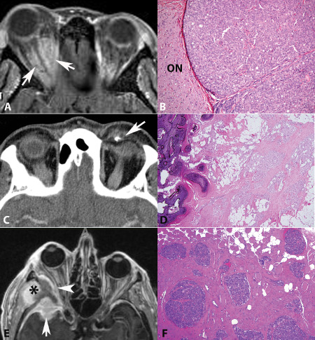
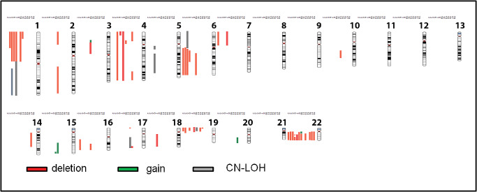
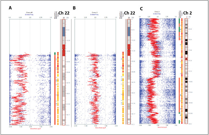
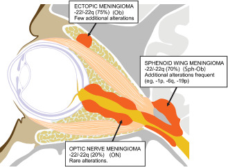
Orbital meningiomas can be classified as primary optic nerve sheath (ON) meningiomas, primary intraorbital ectopic (Ob) meningiomas and spheno-orbital (Sph-Ob) meningiomas based on anatomic site. Single-nucleotide polymorphism (SNP)-based array analysis with the Illumina 300K platform was performed on formalin-fixed, paraffin-embedded tissue from 19 orbital meningiomas (5 ON, 4 Ob and 10 Sph-Ob meningiomas). Tumors were World Health Organization (WHO) grade I except for two grade II meningiomas, and one was NF2-associated. We found genomic alterations in 68% (13 of 19) of orbital meningiomas. Sph-Ob tumors frequently exhibited monosomy 22/22q loss (70%; 7/10) and deletion of chromosome 1p, 6q and 19p (50% each; 5/10). Among genetic alterations, loss of chromosome 1p and 6q were more frequent in clinically progressive tumors. Chromosome 22q loss also was detected in the majority of Ob meningiomas (75%; 3/4) but was infrequent in ON meningiomas (20%; 1/5). In general, Ob tumors had fewer chromosome alterations than Sph-Ob and ON tumors. Unlike Sph-Ob meningiomas, most of the Ob and ON meningiomas did not progress even after incomplete excision, although follow-up was limited in some cases. Our study suggests that ON, Ob and Sph-Ob meningiomas are three molecularly distinct entities. Our results also suggest that molecular subclassification may have prognostic implications.
Keywords: NF2; SNP array; chromosome 22; cytogenetics; optic nerve sheath; orbital meningioma.
© 2014 International Society of Neuropathology.
Figures
Similar articles
-
Diagnostic and prognostic significance of genetic regional heterogeneity in meningiomas.Neuro Oncol. 2004 Oct;6(4):290-9. doi: 10.1215/S1152851704000158. Neuro Oncol. 2004. PMID: 15494096 Free PMC article.
-
NF2 gene mutations and allelic status of 1p, 14q and 22q in sporadic meningiomas.Oncogene. 1999 Apr 1;18(13):2231-9. doi: 10.1038/sj.onc.1202531. Oncogene. 1999. PMID: 10327069
-
Multiple meningioma with different grades of malignancy: case report with genetic analysis applying single-nucleotide polymorphism array and classical cytogenetics.Pathol Res Pract. 2011 Jan 15;207(1):67-72. doi: 10.1016/j.prp.2010.09.001. Pathol Res Pract. 2011. PMID: 20926204
-
Loss of 1p and 7p in radiation-induced meningiomas identified by comparative genomic hybridization.Cancer Genet Cytogenet. 2003 Jul 1;144(1):6-11. doi: 10.1016/s0165-4608(02)00864-6. Cancer Genet Cytogenet. 2003. PMID: 12810249 Review.
-
Pathological classification and molecular genetics of meningiomas.J Neurooncol. 2010 Sep;99(3):379-91. doi: 10.1007/s11060-010-0342-2. Epub 2010 Sep 1. J Neurooncol. 2010. PMID: 20809251 Review.
Cited by
-
Neurofibromatosis type 2 gene mutation and progesterone receptor messenger RNA expression in the pathogenesis of sporadic orbitocranial meningioma.Int J Ophthalmol. 2019 Apr 18;12(4):571-576. doi: 10.18240/ijo.2019.04.07. eCollection 2019. Int J Ophthalmol. 2019. PMID: 31024808 Free PMC article.
-
[Tumors of the inner ear and adjacent structures].Pathologe. 2017 Nov;38(6):521-528. doi: 10.1007/s00292-017-0358-x. Pathologe. 2017. PMID: 28875382 Review. German.
-
Synthesizing Molecular and Immune Characteristics to Move Beyond WHO Grade in Meningiomas: A Focused Review.Front Oncol. 2022 May 31;12:892004. doi: 10.3389/fonc.2022.892004. eCollection 2022. Front Oncol. 2022. PMID: 35712492 Free PMC article. Review.
-
Expressions of Progesterone Receptor of Orbital Meningiomas in Indonesia.Asian Pac J Cancer Prev. 2022 Dec 1;23(12):4137-4143. doi: 10.31557/APJCP.2022.23.12.4137. Asian Pac J Cancer Prev. 2022. PMID: 36579995 Free PMC article.
-
TRAF7 somatic mosaicism in a patient with bilateral optic nerve sheath meningiomas: illustrative case.J Neurosurg Case Lessons. 2022 Jun 6;3(23):CASE2247. doi: 10.3171/CASE2247. eCollection 2022 Jun 6. J Neurosurg Case Lessons. 2022. PMID: 35733823 Free PMC article.
References
-
- Andrews DW, Faroozan R, Yang BP, Hudes RS, Werner‐Wasik M, Kim SM et al (2002) Fractionated stereotactic radiotherapy for the treatment of optic nerve sheath meningiomas: preliminary observations of 33 optic nerves in 30 patients with historical comparison to observation with or without prior surgery. Neurosurgery 51:890–902, discussion 3–4. - PubMed
-
- Bacci C, Sestini R, Provenzano A, Paganini I, Mancini I, Porfirio B et al (2010) Schwannomatosis associated with multiple meningiomas due to a familial SMARCB1 mutation. Neurogenetics 11:73–80. - PubMed
-
- Bosch MM, Wichmann WW, Boltshauser E, Landau K (2006) Optic nerve sheath meningiomas in patients with neurofibromatosis type 2. Arch Ophthalmol 124:379–385. - PubMed
Publication types
MeSH terms
Grants and funding
LinkOut - more resources
Full Text Sources
Other Literature Sources
Miscellaneous
