. 2014 Apr 28;5(2):60.
doi: 10.1186/scrt449.

The effect of Rho kinase inhibition on long-term keratinocyte proliferation is rapid and conditional

The effect of Rho kinase inhibition on long-term keratinocyte proliferation is rapid and conditional

Sandra Chapman et al. Stem Cell Res Ther. .

Abstract

Introduction: We previously demonstrated that the lifespan of primary human keratinocytes could be extended indefinitely by culture in the presence of the Rho kinase (ROCK) inhibitor Y-27632. This technique has proven to be very useful in diverse areas of basic and clinical research.

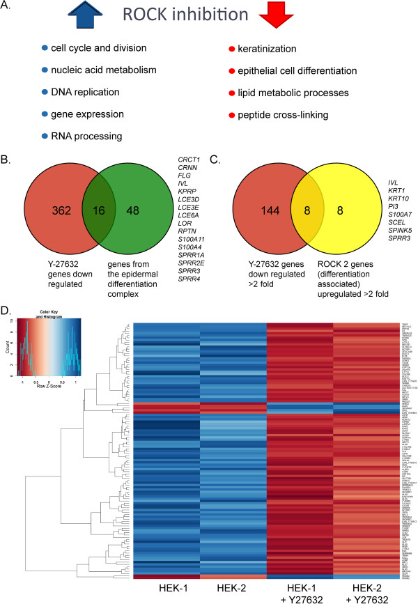
Methods: In this follow-up study we determine whether the continual presence of Y-27632 is required for sustained proliferation. We also test whether different ROCK inhibitors can be used for this technique and whether it can also promote indefinite proliferation of animal keratinocytes. We measure keratinocyte gene expression, proliferation, behaviour and lifespan in the presence and absence of Y-27632.

Results: We demonstrate that the extension of lifespan observed by culture of keratinocytes in the presence of fibroblast feeders and a ROCK inhibitor is reversible and that cells senesce gradually when the inhibitor is removed from the medium. Conversely, keratinocytes that are close to the end of their replicative life span can be revived by ROCK inhibition. We demonstrate that different inhibitors of ROCK can also efficiently extend the lifespan of human keratinocytes and that ROCK inhibition extends the lifespan of animal keratinocytes derived from mouse and bovine epithelia. Gene expression analysis of human epidermal keratinocytes cells grown in the presence of Y-27632 demonstrates that ROCK inhibition primarily inhibits keratinocyte differentiation. Live-imaging of keratinocytes cultured with ROCK inhibitors show that the effect of ROCK inhibition on cellular proliferation is immediate and ROCK inhibited cells proliferate rapidly without differentiation or stratification.

Conclusions: ROCK inhibition rapidly and conditionally induces indefinite proliferation of keratinocytes. This method has far-reaching applications for basic research, as well as for regenerative and personalized medicine.


Figures

Figure 1
Figure 1
Addition and removal of Y-27632 during long-term culture. A. Growth rate of three strains of foreskin keratinocytes from different donors cultured for at least 180 days in the presence or absence of 10 μM Y-27632. Cells cultured with Y-27632 continued to proliferate logarithmically (arrows) while untreated cells senesced at the times indicated by X. B. Y-27632 was removed from the three strains of keratinocytes cultured in its presence at pass 17. The subsequent growth curves are indicated in purple and senescence is indicated by X. C. Y-27632 was removed from the three strains of keratinocytes cultured in its presence at pass 23. The subsequent growth curves are indicated in purple and senescence is indicated by X. D. Y-27632 was added to keratinocyte strain 2 at pass 10. The resulting growth curve is shown in purple. In the inset, all three strains were cultured after cryogenic storage with Y-27632 added at pass 13 or 15 (as indicated). The resulting growth curves are shown. E. Y-27632 was added to keratinocyte strains at pass 19. The resulting growth curves are shown in purple.
Figure 2
Figure 2
Animal keratinocytes proliferate indefinitely in the presence of Y-27632. Growth curves of foetal bovine keratinocytes (A) or new-born mouse keratinocytes (B and C) grown in the absence or presence of Y-27632. Cells in A and C were grown in F-medium in the presence of J2 3T3 feeders. Cells in B were grown in low calcium DMEM in the presence of J2 3T3 feeders. The arrow represents continued proliferation and the X represents senescence. In panel C, the *symbol represents a stage when most cells were senescent; however, a few very rare cells spontaneously transformed and began to proliferate. Panels D, E and F show phase contrast images of bovine (D), mouse (E), and human keratinocytes cultured for the passes shown in the presence of Y-27632. DMEM, Dulbecco’s modified Eagle’s medium.
Figure 3
Figure 3
Effect of different ROCK inhibitors on keratinocyte immortalization. A. Growth curves of the three strains of foreskin keratinocytes cultured in the presence or absence of 10 μM Y-27632, and shown in Figure 1, are reproduced here for comparison with panels B, C and D. B, C, D. Growth curves of the same three strains of foreskin keratinocytes shown in Figures 1 and 2A were cultured in the absence (red) or presence of 20 μM fasudil (B; dark blue line), 20 μM HA-1100 (C, orange line) or 100 nM GSK 429286. (D; light blue line).
Figure 4
Figure 4
Gene expression analysis of human epidermal keratinocytes cultured in the presence or absence of Y-27632. Adult human keratinocytes were cultured for several passes in the presence or absence of 10 μM Y-27632. RNA was isolated at passage 3 and passage 4 and was analysed for gene expression by microarray analysis. A. The major gene ontology (GO) categories up-regulated (blue arrow) and down-regulated (red arrow) are shown. Additional file 2: Table S2 shows the complete list of statistically significantly changed GO categories. B. A comparison of significantly (P <0.05) down-regulated genes from this study with genes encoded by the epidermal differentiation complex [18]. The genes common to both categories are listed to the right. C. A comparison of genes from this study down-regulated at least two-fold by Y-27632 with genes up-regulated by ROCK 2 and associated with keratinocyte differentiation as described by McMullan et al. [19]. The genes common to both categories are listed to the right. D. The microarray data were further analysed for differential gene expression using the R software package Limma [10] and are presented as a heatmap with hierarchical clustering. Down-regulated genes are shown in orange and up-regulated in blue. The list of differentially expressed genes is listed in Additional file 1: Table S1.
Figure 5
Figure 5
Live cell imaging of adult human keratinocytes cultured in the presence or absence of Y-27632. A. Human foreskin keratinocytes at passage 6 were plated in triplicate in the presence or absence of Y-27632. Still images from live movies of each culture condition are shown at the times indicated to represent the confluence of the colonies. The corresponding movies are available as Supplementary data. B. Growth curves were calculated using time-lapse photography to measure confluence of cultures for up to six days (by Incucyte ZOOM software).

References

    1. Chapman S, Liu X, Meyers C, Schlegel R, McBride AA. Human keratinocytes are efficiently immortalized by a Rho kinase inhibitor. J Clin Invest. 2010;120:2619–2626. doi: 10.1172/JCI42297. - DOI - PMC - PubMed
    1. Sakakibara N, Mitra R, McBride AA. The papillomavirus E1 helicase activates a cellular DNA damage response in viral replication foci. J Virol. 2011;85:8981–8995. doi: 10.1128/JVI.00541-11. - DOI - PMC - PubMed
    1. Liu X, Ory V, Chapman S, Yuan H, Albanese C, Kallakury B, Timofeeva OA, Nealon C, Dakic A, Simic V, Haddad BR, Rhim JS, Dritschilo A, Riegel A, McBride AA, Schlegel R. ROCK inhibitor and feeder cells induce the conditional reprogramming of epithelial cells. Am J Pathol. 2012;180:599–607. doi: 10.1016/j.ajpath.2011.10.036. - DOI - PMC - PubMed
    1. Yuan H, Myers S, Wang J, Zhou D, Woo JA, Kallakury B, Ju A, Bazylewicz M, Carter YM, Albanese C, Grant N, Shad A, Dritschilo A, Liu X, Schlegel R. Use of reprogrammed cells to identify therapy for respiratory papillomatosis. N Engl J Med. 2012;367:1220–1227. doi: 10.1056/NEJMoa1203055. - DOI - PMC - PubMed
    1. Jordan CT, Cao L, Roberson ED, Pierson KC, Yang CF, Joyce CE, Ryan C, Duan S, Helms CA, Liu Y, Chen Y, McBride AA, Hwu WL, Wu JY, Chen YT, Menter A, Goldbach-Mansky R, Lowes MA, Bowcock AM. PSORS2 is due to mutations in CARD14. Am J Hum Genet. 2012;90:784–795. doi: 10.1016/j.ajhg.2012.03.012. - DOI - PMC - PubMed

Publication types