. 2014 Apr 28:7:34.
doi: 10.1186/1756-6606-7-34.

Effect of the Brugada syndrome mutation A39V on calmodulin regulation of Cav1.2 channels

Affiliations

Effect of the Brugada syndrome mutation A39V on calmodulin regulation of Cav1.2 channels

Brett A Simms et al. Mol Brain. .

Abstract

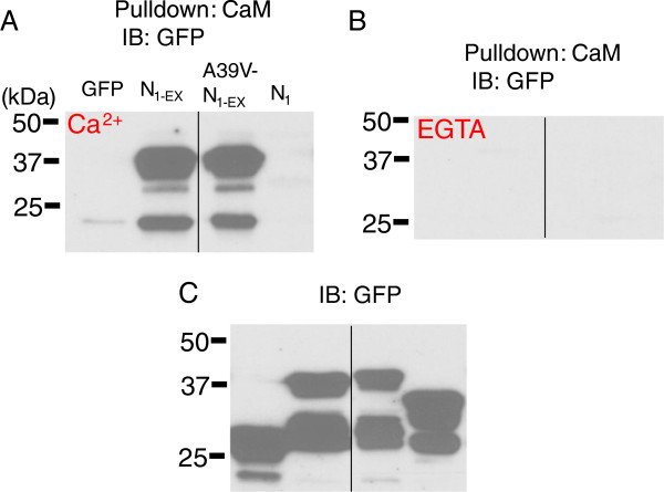
Background: The L-type calcium channel Cav1.2 is important for brain and heart function. The ubiquitous calcium sensing protein calmodulin (CaM) regulates calcium dependent gating of Cav1.2 channels by reducing calcium influx, a process known as calcium-dependent inactivation (CDI). Dissecting the calcium-dependence of CaM in this process has benefited greatly from the use of mutant CaM molecules which are unable to bind calcium to their low affinity (N-lobe) and high affinity (C-lobe) binding sites. Unlike CDI, it is unknown whether CaM can modulate the activation gating of Cav1.2 channels.

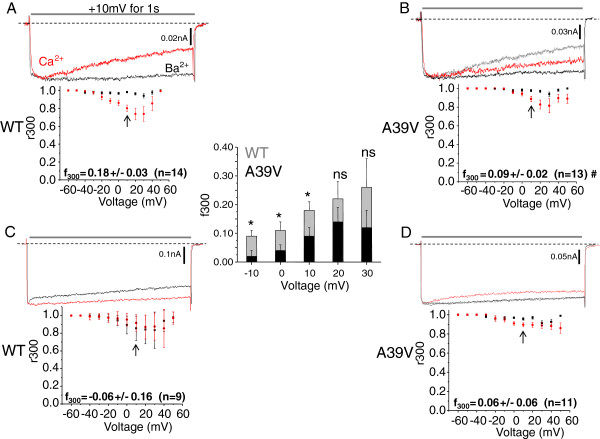
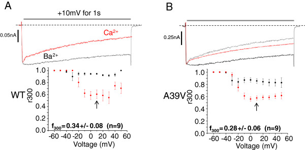
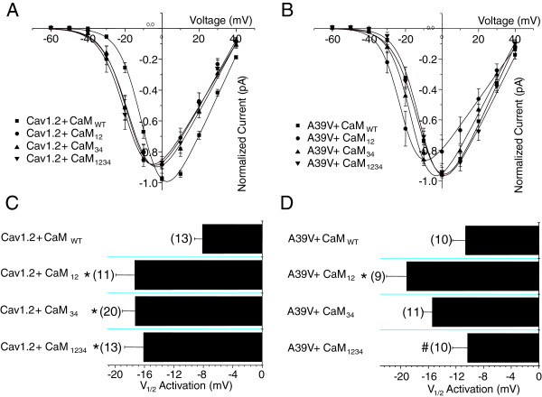
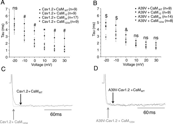
Results: We examined a Cav1.2 point mutant in the N-terminus region of the channel (A39V) that has been previously linked to Brugada syndrome. Using mutant CaM constructs in which the N- and/or C-lobe calcium binding sites were ablated, we were able to show that this Brugada syndrome mutation disrupts N-lobe CDI of the channel. In the course of these experiments, we discovered that all mutant CaM molecules were able to alter the kinetics of channel activation even in the absence of calcium for WT-Cav1.2, but not A39V-Cav1.2 channels. Moreover, CaM mutants differentially shifted the voltage-dependence of activation for WT and A39V-Cav1.2 channels to hyperpolarized potentials. Our data therefore suggest that structural changes in CaM that arise directly from site directed mutagenesis of calcium binding domains alter activation gating of Cav1.2 channels independently of their effects on calcium binding, and that the N-terminus of the channel contributes to this CaM dependent process.

Conclusions: Our data indicate that caution must be exercised when interpreting the effects of CaM mutants on ion channel gating.


Figures

Figure 1
Figure 1
The Brugada syndrome mutation A39V disrupts N-lobe CDI of Cav1.2 channels. A) Representative Ba2+ (black) and Ca2+ (red) traces of WT and A39V-Cav1.2 channels (B) expressed with Cavβ2a/Cavα2δ in the presence of low calcium buffering (0.5 mM EGTA) and CaM34. Note that the peak of the Ba2+ trace is normalized to that in the presence of Ca2+. For reference the WT-Cav1.2 Ca2+ trace is displayed in grey in (B). The plots shown below the current traces reflect average CDI (f300) which is quantified by the fraction of current remaining after 300 ms (r300) in calcium, and is then subtracted from the fraction of current remaining in barium at the same time point. The f300 value at 10 mV (arrows) is significantly less for A39V, than WT-Cav1.2 (# p ≤ 0.04 by student’s t-test). The inset bar graph displays additional f300 values over a potential range from -10 mV to +30 mV. A significant difference is observed in the f300 values between WT-Cav1.2 and A39V-Cav1.2 at −10, 0 and +10 mV (p ≤ 0.05 by student’s t-test), but not at +20 (p = 0.28 by student’s t-test) or +30 mV (p = 0.26 by student’s t-test). C) Under high calcium buffering (10 mM BAPTA) conditions WT-Cav1.2 channels expressed as in (A) no longer exhibit N-lobe CDI. D) A39V-Cav1.2 does not show significant N-lobe CDI compared to WT-Cav1.2 in high calcium buffering at +10 mV (arrows). For reference the WT-Cav1.2 Ca2+ trace (C) is displayed in grey.
Figure 2
Figure 2
The Brugada syndrome mutation A39V does not affect C-lobe CDI of Cav1.2 channels. A) Representative Ba2+ (black) and Ca2+ (red) traces of WT-Cav1.2 channels expressed with Cavβ2a/Cavα2δ and CaM12 in the presence of high calcium buffering (10 mM BAPTA). Note that the peak of the Ba2+ trace is normalized to that in the presence of Ca2+ and that average CDI (f300) of WT-Cav1.2 is displayed below in the graph. B) A39V-Cav1.2 channels expressed with Cavβ2a/Cavα2δ and CaM12 in the presence of high calcium buffering (10 mM BAPTA). Inset graphs show average CDI (f300) which is not statistically different (p ≤ 0.55 by student’s t-test) between the two channel types.
Figure 3
Figure 3
The Brugada mutation A39V does not alter N-terminal binding to CaM. A) CaM sepharose pull-down experiments of N1-EX, A39V-N1-EX and N1 GFP fusion proteins in 0.5 mM Ca2+, or 5 mM EGTA (B) run on SDS-PAGE with corresponding lysates (C) and blotted for GFP. Black lines mark where the gel picture was cut and irrelevant samples removed. These experiments were performed twice each.
Figure 4
Figure 4
CaM lobe mutants differentially shift the voltage dependence of activation for WT and A39V-Cav1.2 channels. A) Current voltage relationships for WT-Cav1.2 channels expressed transiently in tsA-201 cells with Cavβ2a/Cavα2δ and recorded in barium with one of four CaM conditions: CaMWT, CaM12, CaM34, or CaM1234. All experiments were recorded with high calcium buffering intracellularly (10 mM BAPTA). B) Current–voltage relationships for A39V-Cav1.2 channels expressed as in (A) with one of four CaM conditions: CaMWT, CaM12, CaM34, or CaM1234. C) A bar graph displaying the half activation potentials for Cav1.2 channels recorded in barium. WT-Cav1.2 channels recorded with any CaM mutant have a significant leftward shift in the voltage-dependence of activation in barium compared to CaMWT (*p ≤ 0.05 by one-way ANOVA). D) A bar graph displaying the voltage dependence of activation for A39V-Cav1.2 channels recorded in barium. A39V-Cav1.2 channels recorded with CaM12 have a significant leftward shift in the voltage-dependence of activation in barium compared to CaMWT (*p ≤ 0.05 by one-way ANOVA), and CaM1234 (# p ≤ 0.05 by one-way ANOVA).
Figure 5
Figure 5
Calmodulin lobe mutants differentially affect the kinetics of activation for WT and A39V-Cav1.2 channels in the absence of calcium. A) Plot illustrating the time to maximum activation (Tau) at various voltages for WT-Cav1.2 channels expressed transiently in tsA-201 cells with Cavβ2a/Cavα2δ and recorded in barium with one of CaMWT, CaM12, CaM34 or CaM1234 and with 10 mM BAPTA intracellular. Voltages for which all mutant CaMs differ significantly from the CaMWT condition (* p ≤ 0.05 by one-way ANOVA), and where CaM34 and CaM1234 differ from CaMWT condition (# p ≤ 0.05 by one-way ANOVA). B) Plot for the kinetics of activation of A39V-Cav1.2 channels expressed as in (A). Voltages for which CaM1234 differs significantly from the CaMWT condition ($ p ≤ 0.05 by one-way ANOVA), and where CaM12 and CaM1234 differ from CaMWT condition (& p ≤ 0.05 by one-way ANOVA). C) Sample traces of WT-Cav1.2 channels expressed with CaMWT and CaM1234 at 10 mV. Note that the CaM1234 trace has been normalized to that of CaMWT and that the arrows denote peak of activation for WT-Cav1.2 with either CaMWT (black) or CaM1234 (grey). D) Sample traces of A39V-Cav1.2 channels expressed with CaMWT and CaM1234 at 10 mV. Note that the CaM1234 trace has been normalized to that of CaMWT and that the arrows denote peak of activation for A39V-Cav1.2 with either CaMWT (black) or CaM1234 (grey).

References

    1. Catterall WA. Structure and regulation of voltage-gated Ca2+ channels. Annu Rev Cell Dev Biol. 2000;16:521–551. doi: 10.1146/annurev.cellbio.16.1.521. - DOI - PubMed
    1. Simms BA, Zamponi GW. Neuronal voltage-gated calcium channels: structure, function and dysfunction. Neuron. 2014;82:24–45. doi: 10.1016/j.neuron.2014.03.016. - DOI - PubMed
    1. Jeon D, Kim S, Chetana M, Jo D, Ruley HE, Lin SY, Rabah D, Kinet JP, Shin HS. Observational fear learning involves affective pain system and Cav1.2 Ca2+ channels in ACC. Nat Neurosci. 2010;13(4):482–488. doi: 10.1038/nn.2504. - DOI - PMC - PubMed
    1. Langwieser N, Christel CJ, Kleppisch T, Hofmann F, Wotjak CT, Moosmang S. Homeostatic switch in hebbian plasticity and fear learning after sustained loss of Cav1.2 calcium channels. J Neurosci. 2012;30(25):8367–8375. - PMC - PubMed
    1. Shaw RM, Colecraft HM. L-type calcium channel targeting and local signalling in cardiac myocytes. Cardiovasc Res. 2013;98(2):177–186. doi: 10.1093/cvr/cvt021. - DOI - PMC - PubMed

Publication types

LinkOut - more resources