. 2014 Apr 28;15(1):54.
doi: 10.1186/1465-9921-15-54.

Atrial fibrosis in a chronic murine model of obstructive sleep apnea: mechanisms and prevention by mesenchymal stem cells

Affiliations

Atrial fibrosis in a chronic murine model of obstructive sleep apnea: mechanisms and prevention by mesenchymal stem cells

Pablo Ramos et al. Respir Res. .

Abstract

Background: OSA increases atrial fibrillation (AF) risk and is associated with poor AF treatment outcomes. However, a causal association is not firmly established and the mechanisms involved are poorly understood. The aims of this work were to determine whether chronic obstructive sleep apnea (OSA) induces an atrial pro-arrhythmogenic substrate and to explore whether mesenchymal stem cells (MSC) are able to prevent it in a rat model of OSA.

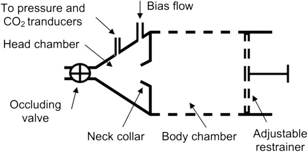
Methods: A custom-made setup was used to mimic recurrent OSA-like airway obstructions in rats. OSA-rats (n = 16) were subjected to 15-second obstructions, 60 apneas/hour, 6 hours/day during 21 consecutive days. Sham rats (n = 14) were placed in the setup but no obstructions were applied. In a second series of rats, MSC were administered to OSA-rats and saline to Sham-rats. Myocardial collagen deposit was evaluated in Picrosirius-red stained samples. mRNA expression of genes involved in collagen turnover, inflammation and oxidative stress were quantified by real time PCR. MMP-2 protein levels were quantified by Western Blot.

Results: A 43% greater interstitial collagen fraction was observed in the atria, but not in the ventricles, of OSA-rats compared to Sham-rats (Sham 8.32 ± 0.46% vs OSA 11.90 ± 0.59%, P < 0.01). Angiotensin-I Converting Enzyme (ACE) and Interleukin 6 (IL-6) expression were significantly increased in both atria, while Matrix Metalloproteinase-2 (MMP-2) expression was decreased. MSC administration blunted OSA-induced atrial fibrosis (Sham + Saline 8.39 ± 0.56% vs OSA + MSC 9.57 ± 0.31%, P = 0.11), as well as changes in MMP-2 and IL-6 expression. Interleukin 1-β (IL-1β) plasma concentration correlated to atrial but not ventricular fibrosis. Notably, a 2.5-fold increase in IL-1β plasma levels was observed in the OSA group, which was prevented in rats receiving MSC.

Conclusions: OSA induces selective atrial fibrosis in a chronic murine model, which can be mediated in part by the systemic and local inflammation and by decreased collagen-degradation. MSCs transplantation prevents atrial fibrosis, suggesting that these stem cells could counterbalance inflammation in OSA.


Figures

Figure 1
Figure 1
Experimental obstructive sleep apnea (OSA) setup. Diagram of the experimental setup to noninvasively apply recurrent airway obstructions in the rat. See text for explanation.
Figure 2
Figure 2
Fibrosis assessment in OSA and Sham rats. A. Representative Picrosirius-stained photomicrographs of atrial, right ventricle (RV) and left ventricle (LV) sections from Sham (n = 14) and OSA (n = 16) rats. Atria and ventricular samples were stained in different days, and pictures obtained at different magnifications, so results from atria and ventricles should not be compared. B. Quantification of collagen fraction in the atria, RV and LV. (Mean ± SEM, *P < 0.05).
Figure 3
Figure 3
Fibrosis mechanisms assessment in OSA and Sham rats. (A) ACE, (B) MMP-2 and (C) IL-6 mRNA expression (ng-equ) in the four cardiac chambers of Sham (n = 14) and OSA (n = 16) rats (Mean ± SEM, *P < 0.05). D. Plasmatic concentration (pg/mL) of IL-1β in Sham and OSA (Mean ± SEM, *P < 0.05). E. MMP-2 protein levels quantification. Representative MMP-2 blots (upper panel), Ponceau-stained membranes (middle panel) and normalized quantification (lower panel) (Mean ± SEM, *P < 0.05). n = 4-6/group. Left lane is a molecular-weight marker lane; the picture was obtained with visible light. Dashed line means non-contiguous lane. A.U.: Arbitrary Units.
Figure 4
Figure 4
mRNA expression in the OSA and Sham rats. (A) TIMP-1, (B) TIMP-2, (C) MMP-9, (D) LOX, (E) eNOS and (F) TGF-β1 mRNA expression (ng-equ) in the four cardiac chambers of Sham (n = 14) and OSA (n = 16) rats (Mean ± SEM, *P < 0.05).
Figure 5
Figure 5
Correlation between plasma IL-1β and myocardial fibrosis at the atrial (A) and ventricular (B) level. Dashed line means 95% confidence interval.
Figure 6
Figure 6
Fibrosis assessment in OSA + MSC and Sham + S rats. A. Picrosirius-stained photomicrographs of atrial sections, right ventricular (RV) sections and left ventricular (LV) sections from Sham + Saline (n = 7) and OSA + MSC (n = 8) rats. Atria and ventricular samples were stained in different days, and pictures obtained at different magnification, so results from atria and ventricles should not be compared. B. Quantification of collagen fraction in the atria, RV and LV measured in Picrosirius-red stained samples (Mean ± SEM).
Figure 7
Figure 7
CD90 immunofluorescence myocardial stained samples. A. Cultured MSC, positive control for CD90 (40x). 100x augmentation in the upper box. Abundant CD90 deposits (some marked with arrowhead) are present. B. Cultured MSC, negative control (x40, no anti-CD90 antibody added). C. Sample of interest (left atrium), CD90 stained. No positive cells are marked (x40). D. Sample of interest, negative control (x40, no anti-CD90 antibody added). Blue: nuclei (DAPI); green: CD90.
Figure 8
Figure 8
Assessment of fibrosis mechanisms in OSA + MSC vs Sham + S rats. (A) ACE, (B) MMP-2 and (C) IL-6 mRNA expression (ng-equ) in the four cardiac chambers of Sham + Saline (n = 7) and OSA + MSC (n = 8) rats (Mean ± SEM *P < 0.05). D. Plasmatic concentration (pg/mL) of IL-1β in Sham + Saline and OSA + MSC rats (Mean ± SEM *P < 0.05). E. MMP-2 protein levels quantification. Representative MMP-2 blots (upper panel), Ponceau-stained membranes (middle panel) and normalized quantification (lower panel) (Mean ± SEM). n = 4-6/group. Left lane is a molecular-weight marker lane; the picture was obtained with visible light. A.U.: Arbitrary Units.
Figure 9
Figure 9
mRNA expression in the OSA + MSC and Sham + S rats. (A) TIMP-1, (B) TIMP-2, (C) MMP-9, (D) LOX, (E) eNOS and (F) TGF-β1 mRNA expression (ng-equ) in the four cardiac chambers of Sham + Saline (n = 7) and OSA + MSC (n = 8) rats (Mean ± SEM *P < 0.05).
Figure 10
Figure 10
Proposed pathophysiology of OSA-induced atrial fibrosis and MSC mechanism of action. OSA acts as a pro-inflammatory stimulus and inhibits MMP-2 synthesis, reducing collagen degradation and thus favoring collagen accumulation. MSC administration blunts inflammation and normalizes MMP-2 synthesis, thereby increasing collagen degradation and preventing from collagen deposit. OSA also increases ACE expression, but its role in fibrosis promotion is uncertain. Red lines represent inhibition, green lines represent activation. MSC: mesenchymal stem cells. OSA: obstructive sleep apnea.

References

    1. Gami AS, Pressman G, Caples SM, Kanagala R, Gard JJ, Davison DE, Malouf JF, Ammash NM, Friedman PA, Somers VK. Association of atrial fibrillation and obstructive sleep apnea. Circulation. 2004;110:364–367. doi: 10.1161/01.CIR.0000136587.68725.8E. - DOI - PubMed
    1. Gami AS, Hodge DO, Herges RM, Olson EJ, Nykodym J, Kara T, Somers VK. Obstructive sleep apnea, obesity, and the risk of incident atrial fibrillation. J Am Coll Cardiol. 2007;49:565–571. doi: 10.1016/j.jacc.2006.08.060. - DOI - PubMed
    1. Kanagala R, Murali NS, Friedman PA, Ammash NM, Gersh BJ, Ballman KV, Shamsuzzaman AS, Somers VK. Obstructive sleep apnea and the recurrence of atrial fibrillation. Circulation. 2003;107:2589–2594. - PubMed
    1. Matiello M, Nadal M, Tamborero D, Berruezo A, Montserrat J, Embid C, Rios J, Villacastin J, Brugada J, Mont L. Low efficacy of atrial fibrillation ablation in severe obstructive sleep apnoea patients. Europace. 2010;12:1084–1089. doi: 10.1093/europace/euq128. - DOI - PubMed
    1. Ng CY, Liu T, Shehata M, Stevens S, Chugh SS, Wang X. Meta-analysis of obstructive sleep apnea as predictor of atrial fibrillation recurrence after catheter ablation. Am J Cardiol. 2011;108:47–51. doi: 10.1016/j.amjcard.2011.02.343. - DOI - PubMed

Publication types