. 2014 Jul;58(7):3860-6.
doi: 10.1128/AAC.02064-13. Epub 2014 Apr 28.

The broad-spectrum antiviral compound ST-669 restricts chlamydial inclusion development and bacterial growth and localizes to host cell lipid droplets within treated cells

Affiliations

The broad-spectrum antiviral compound ST-669 restricts chlamydial inclusion development and bacterial growth and localizes to host cell lipid droplets within treated cells

Kelsi M Sandoz et al. Antimicrob Agents Chemother. 2014 Jul.

Abstract

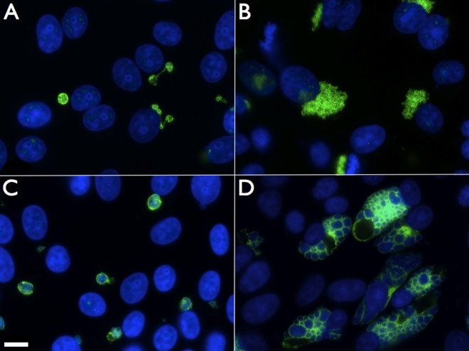
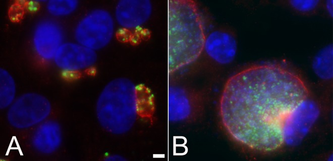
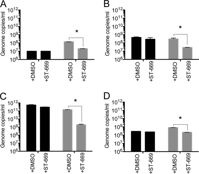
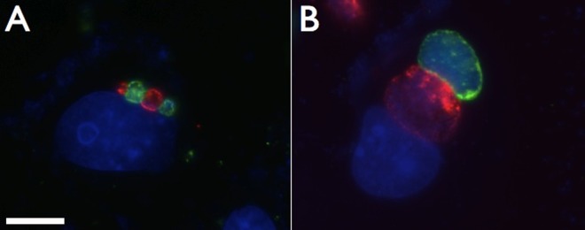
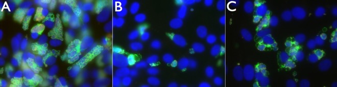
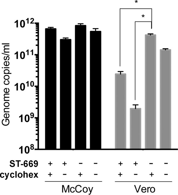
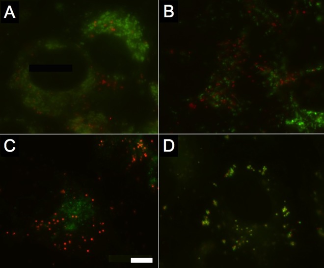
Novel broad-spectrum antimicrobials are a critical component of a strategy for combating antibiotic-resistant pathogens. In this study, we explored the activity of the broad-spectrum antiviral compound ST-669 for activity against different intracellular bacteria and began a characterization of its mechanism of antimicrobial action. ST-669 inhibits the growth of three different species of chlamydia and the intracellular bacterium Coxiella burnetii in Vero and HeLa cells but not in McCoy (murine) cells. The antichlamydial and anti-C. burnetii activity spectrum was consistent with those observed for tested viruses, suggesting a common mechanism of action. Cycloheximide treatment in the presence of ST-669 abrogated the inhibitory effect, demonstrating that eukaryotic protein synthesis is required for tested activity. Immunofluorescence microscopy demonstrated that different chlamydiae grow atypically in the presence of ST-669, in a manner that suggests the compound affects inclusion formation and organization. Microscopic analysis of cells treated with a fluorescent derivative of ST-669 demonstrated that the compound localized to host cell lipid droplets but not to other organelles or the host cytosol. These results demonstrate that ST-669 affects intracellular growth in a host-cell-dependent manner and interrupts proper development of chlamydial inclusions, possibly through a lipid droplet-dependent process.


Figures

FIG 1
FIG 1
ST-669 treatment alters the size and structure of C. caviae inclusions. Immunofluorescence microscopy of ST-669 (A and C)- and DMSO (B and D)-treated C. caviae-infected Vero cells fixed at 20 h (A and B) or 40 h (C and D) postinfection was performed. Images were taken at ×100 magnification, and the scale bar in panel C indicates 10 μm for each panel. C. caviae inclusions were labeled with anti-IncA (green), and total DNA was labeled with DAPI (blue).
FIG 2
FIG 2
Treatment of C. trachomatis-infected Vero cells with ST-669 affects inclusion size and chlamydial abundance. Infected and treated Vero cells were fixed with methanol at 40 h postinfection. Fixed monolayers were labeled with anti-IncA (red), anti-MOMP (green), and DAPI for DNA (blue) and visualized using a ×100 objective lens. Infected cells were treated with either ST-669 (A) or the vehicle, DMSO (B). The scale bar in panel A indicates 5 μm for both panels.
FIG 3
FIG 3
ST-669 inhibits growth of Coxiella burnetii and Chlamydia spp. in a host-specific manner. qPCR analyses of ST-669- and DMSO-treated Coxiella burnetii (A; 72 h postinfection), C. muridarum (B; 20 h postinfection), C. trachomatis L2 (C; 40 h postinfection), and C. caviae GPIC (D; 40 h postinfection) inside McCoy (black) or Vero (gray) cells. All experiments were conducted at an MOI of 1. Genome copies/ml are plotted on the y axis. The asterisk indicates statistically significant differences (P < 0.0001).
FIG 4
FIG 4
Coinfection of C. caviae and C. trachomatis demonstrate the altered inclusion phenotype of ST-669-treated C. caviae. Vero cells treated with 10 μM ST-669 were coinfected with C. caviae (green) and C. trachomatis (red), followed by incubation for 20 h (A) and 50 h (B). The cells were then fixed and labeled with species-specific anti-IncA antibodies. The total DNA was labeled with DAPI (blue). The scale bar in panel A indicates 10 μm for each panel.
FIG 5
FIG 5
Cycloheximide and ST-669 cotreatment of C. caviae-infected cells. Vero cells were infected with C. caviae and treated with either DMSO plus cycloheximide (A), ST-669 (B), or ST-669 plus cycloheximide (C). The cells were then cultured for 40 h, fixed with methanol, and stained for immunofluorescence microscopy. Images were visualized for total DNA (blue), chlamydial LPS (red), and IncA (green), using the ×40 objective lens.
FIG 6
FIG 6
Cycloheximide treatment partially reverses the inhibitory effect of ST-669. qPCR of ST-669-treated or untreated (i.e., DMSO only) C. trachomatis L2-infected McCoy and Vero cells in the presence or absence of 100 μg of cycloheximide (cyclohex)/ml. Infections were sonicated, and gDNA was collected at 40 h postinfection before assay by qPCR. The asterisk indicates statistically significant differences (P < 0.0001).
FIG 7
FIG 7
Dual labeling of host cell structures with ST-669F shows that ST-669F trafficks to intracellular lipid droplets. Uninfected Vero cells were labeled with ST-669F (red) and probes specific to different organelles (green) to determine colocalization of the ST-669F molecule. (A) Lysosomes; (B) mitochondria; (C) Golgi apparatus; (D) lipid droplets. The scale bar in panel C indicates 5 μm for each panel.
FIG 8
FIG 8
Colocalization of ST-669F and Bodipy 493/503 in both McCoy and Vero cells treated with oleic acid. Uninfected McCoy cells (top row) and Vero cells (bottom row) were treated with 100 μM oleic acid for 16 h and then labeled with a combination of ST-669F (green) and Bodipy 493/503 (red) to determine colocalization of the ST-669F molecule. Bodipy 493/503 and ST-669F were added to the culture medium as indicated in Materials and Methods, and then the cells were visualized for each label. (A and D) Bodipy 493/503-labeled lipid droplets; (B and E) ST-669F, pseudocolored green; (C and F) merged image of each of the two panels.

References

    1. Starnbach MN, Roan NR. 2008. Conquering sexually transmitted diseases. Nat. Rev. Immunol. 8:313–317. 10.1038/nri2272 - DOI - PubMed
    1. Abdelrahman YM, Belland RJ. 2005. The chlamydial developmental cycle. FEMS Microbiol. Rev. 29:949–959. 10.1016/j.femsre.2005.03.002 - DOI - PubMed
    1. Carabeo RA, Mead DJ, Hackstadt T. 2003. Golgi-dependent transport of cholesterol to the Chlamydia trachomatis inclusion. Proc. Natl. Acad. Sci. U. S. A. 100:6771–6776. 10.1073/pnas.1131289100 - DOI - PMC - PubMed
    1. Hatch GM, McClarty G. 1998. Phospholipid composition of purified Chlamydia trachomatis mimics that of the eucaryotic host cell. Infect. Immun. 66:3727–3735 - PMC - PubMed
    1. Moore ER, Fischer ER, Mead DJ, Hackstadt T. 2008. The chlamydial inclusion preferentially intercepts basolaterally directed sphingomyelin-containing exocytic vacuoles. Traffic 9:2130–2140. 10.1111/j.1600-0854.2008.00828.x - DOI - PMC - PubMed

Publication types

LinkOut - more resources