. 2014 Jul;58(7):3914-20.
doi: 10.1128/AAC.02363-13. Epub 2014 Apr 28.

Crystal structure of Mox-1, a unique plasmid-mediated class C β-lactamase with hydrolytic activity towards moxalactam

Affiliations

Crystal structure of Mox-1, a unique plasmid-mediated class C β-lactamase with hydrolytic activity towards moxalactam

Takuma Oguri et al. Antimicrob Agents Chemother. 2014 Jul.

Abstract

Mox-1 is a unique plasmid-mediated class C β-lactamase that hydrolyzes penicillins, cephalothin, and the expanded-spectrum cephalosporins cefepime and moxalactam. In order to understand the unique substrate profile of this enzyme, we determined the X-ray crystallographic structure of Mox-1 β-lactamase at a 1.5-Å resolution. The overall structure of Mox-1 β-lactamase resembles that of other AmpC enzymes, with some notable exceptions. First, comparison with other enzymes whose structures have been solved reveals significant differences in the composition of amino acids that make up the hydrogen-bonding network and the position of structural elements in the substrate-binding cavity. Second, the main-chain electron density is not observed in two regions, one containing amino acid residues 214 to 216 positioned in the Ω loop and the other in the N terminus of the B3 β-strand corresponding to amino acid residues 303 to 306. The last two observations suggest that there is significant structural flexibility of these regions, a property which may impact the recognition and binding of substrates in Mox-1. These important differences allow us to propose that the binding of moxalactam in Mox-1 is facilitated by the avoidance of steric clashes, indicating that a substrate-induced conformational change underlies the basis of the hydrolytic profile of Mox-1 β-lactamase.


Figures

FIG 1
FIG 1
Chemical structures of cephalothin and moxalactam.
FIG 2
FIG 2
Multiple-sequence alignment of class C β-lactamases whose structures have been solved. The sequences of the structure-solved class C enzymes that have high similarity with Mox-1 were aligned by use of the DaliLite (v3) program (20). The aligned enzymes (with their Protein Databank and Uniprot accession numbers) are as follows: Mox-1 (Uniprot accession number Q51578); AmpC from P. aeruginosa PAO1 (PDB accession no. 4GZB, Uniprot accession no. P24735); AmpC from Pseudomonas fluorescens (PFLU; PDB accession no. 2QZ6, Uniprot accession no.P85302); AmpC from E. cloacae P99 (PDB accession no. 1XX2, Uniprot accession no. P05364); GC1, which is an AmpC enzyme from E. cloacae (PDB accession no. 1GCE, Uniprot accession no. Q59401); CMY-2 (PDB accession no. 1ZC2, Uniprot accession no. Q48434); and AmpC from E. coli (ECOLI; PDB accession no. 1KE4, Uniprot accession no. P00811). The residue different between Mox-1 and CMY-10 is marked with a black dot. Residues that are conserved in all the enzymes listed here are shown with white letters with a black background. One of the three conserved regions is boxed. The Mox-1 residues whose structural model is not built because of a lack of electron density are shown in lowercase. The residues mentioned in the text are indicated with arrows and the residue name and number. The secondary structure of Mox-1 mentioned in the text is shown just below its sequence.
FIG 3
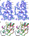
FIG 3
The active site of Mox-1. (A) Stereoview of the active site 2FoFc electron density map contoured at 1.5 σ. The hydrogen-bonding interaction is shown with a dotted line. (B) The active site of Mox-1 in comparison with that of other class C enzymes whose structures have been solved. The backbone and side-chain carbons of Mox-1 (green), CMY-10 (cyan), AmpC from P. aeruginosa (yellow), P99 (magenta), GC1 (pale pink), and AmpC from E. coli (orange) are shown.
FIG 4
FIG 4
Disordered B2g-B3 loop in Mox-1. The region containing residues 303 to 306 of Mox-1 is shown in green, and the corresponding regions of CMY-10, AmpC from P. aeruginosa, and P99 are shown in cyan, magenta, and pale pink, respectively, on the molecular surface of Mox-1. The disordered region is indicated with a dotted line and black arrow. The active-site cavity is shown with a red arrow.
FIG 5
FIG 5
Superposition of Mox-1 and the E. coli AmpC enzyme complexed with moxalactam. Mox-1 carbon atoms are shown in green. Two molecules of E. coli AmpC enzymes complexed with moxalactam (molecules A and B in the protein with PDB accession no. 1FCO) are shown in pale orange and pale pink, respectively, and the moxalactam molecules bound to them are shown in magenta and purple, respectively.

References

    1. Wilke MS, Lovering AL, Strynadka NC. 2005. β-Lactam antibiotic resistance: a current structural perspective. Curr. Opin. Microbiol. 8:525–533. 10.1016/j.mib.2005.08.016 - DOI - PubMed
    1. Ambler RP. 1980. The structure of β-lactamases. Philos. Trans. R. Soc. Lond. B Biol. Sci. 289:321–331. 10.1098/rstb.1980.0049 - DOI - PubMed
    1. Bulychev A, Mobashery S. 1999. Class C β-lactamases operate at the diffusion limit for turnover of their preferred cephalosporin substrates. Antimicrob. Agents Chemother. 43:1743–1746 - PMC - PubMed
    1. Bush K, Jacoby GA, Medeiros AA. 1995. A functional classification scheme for β-lactamases and its correlation with molecular structure. Antimicrob. Agents Chemother. 39:1211–1233. 10.1128/AAC.39.6.1211 - DOI - PMC - PubMed
    1. Jacoby GA. 2009. AmpC β-lactamases. Clin. Microbiol. Rev. 22:161–182. 10.1128/CMR.00036-08 - DOI - PMC - PubMed

Publication types

MeSH terms

Associated data

LinkOut - more resources