. 2014 Jul;34(14):2611-23.
doi: 10.1128/MCB.01121-13.

Caveolin-1 regulates genomic action of the glucocorticoid receptor in neural stem cells

Caveolin-1 regulates genomic action of the glucocorticoid receptor in neural stem cells

Melanie E Peffer et al. Mol Cell Biol. 2014 Jul.

Abstract

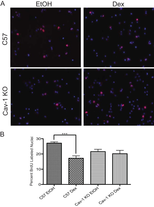
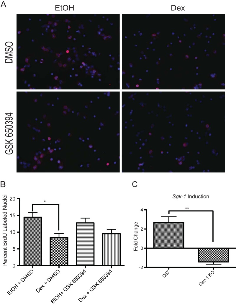
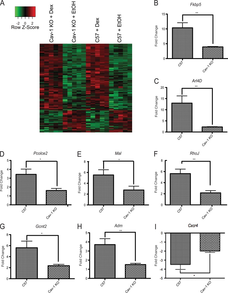
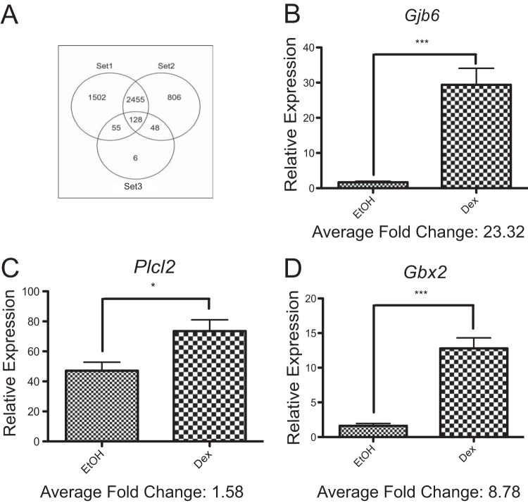
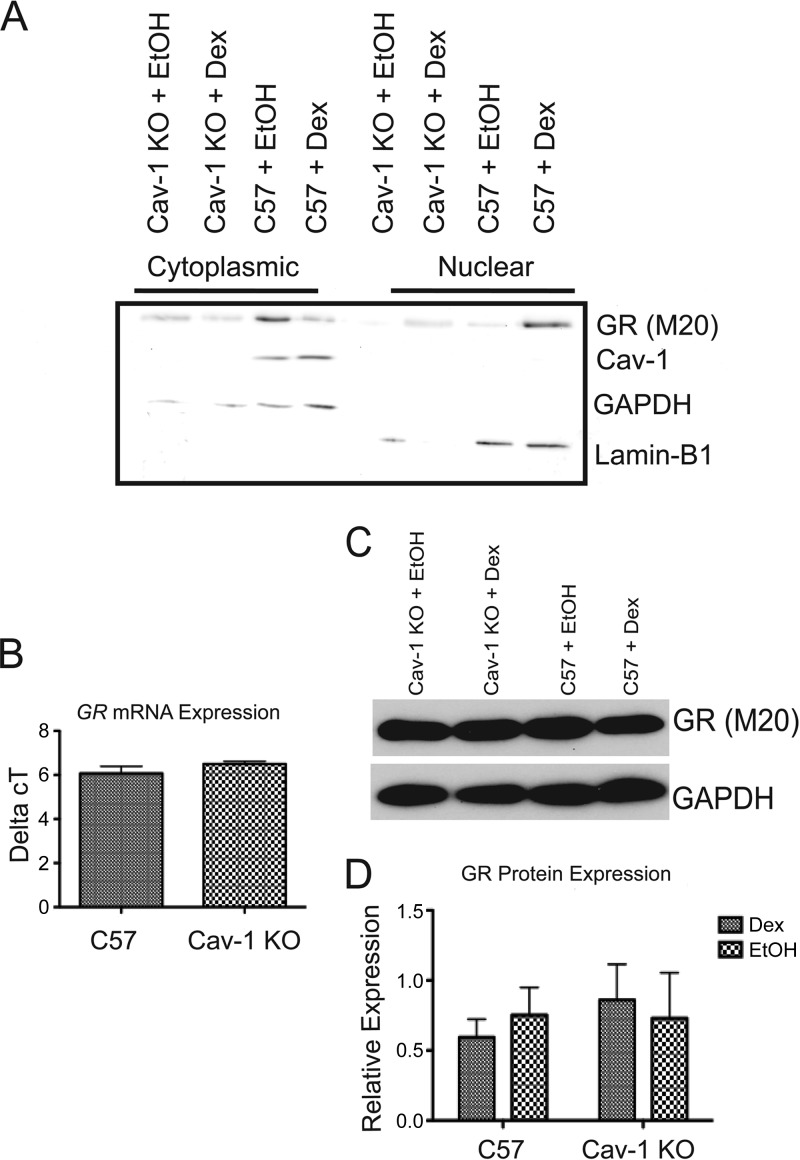
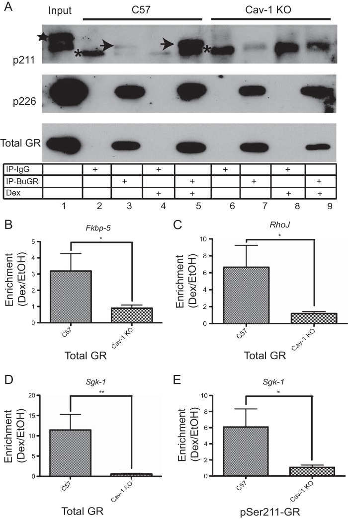
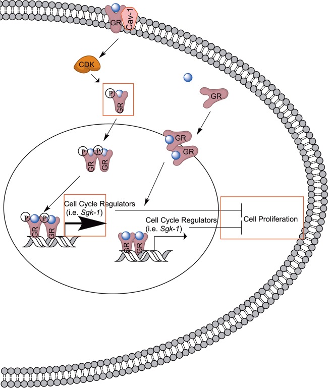
While glucocorticoids (GCs) are used clinically to treat many conditions, their neonatal and prenatal usage is increasingly controversial due to reports of delayed adverse outcomes, especially their effects on brain development. Such alterations may reflect the impact of GCs on neural progenitor/stem cell (NPSC) function. We previously demonstrated that the lipid raft protein caveolin-1 (Cav-1) was required for rapid GC signaling in embryonic mouse NPSCs operating through plasma membrane-bound glucocorticoid receptors (GRs). We show here that genomic GR signaling in NPSCs requires Cav-1. Loss of Cav-1 impacts the transcriptional response of many GR target genes (e.g., the serum- and glucocorticoid-regulated kinase 1 gene) that are likely to mediate the antiproliferative effects of GCs. Microarray analysis of wild-type C57 or Cav-1-deficient NPSCs identified approximately 100 genes that are differentially regulated by GC treatment. These changes in hormone responsiveness in Cav-1 knockout NPSCs are associated with the loss of GC-regulated phosphorylation of GR at serine 211 but not at serine 226. Chromatin recruitment of total GR to regulatory regions of target genes such as Fkbp-5, RhoJ, and Sgk-1, as well as p211-GR recruitment to Sgk-1, are compromised in Cav-1 knockout NPSCs. Cav-1 is therefore a multifunctional regulator of GR in NPSCs influencing both rapid and genomic action of the receptor to impact cell proliferation.


Figures

FIG 1
FIG 1
Antiproliferative effects of Dex in NPSCs require Cav-1. (A) C57 or Cav-1 KO NPSCs were treated with Dex or vehicle (ethanol [EtOH]) for 24 h and pulsed with BrdU during the last hour of treatment. Immunocytochemistry was performed to detect BrdU-positive nuclei (pink). Nuclei were visualized by DAPI staining (blue). (B) Three independent coverslips per biological replicate were counted to ascertain the percentage of cells that passed through S-phase (i.e., BrdU-positive nuclei). Error bars represent the standard error of the mean (SEM; n = three biological replicates). P < 0.001 (one-way ANOVA with Tukey's posttest).
FIG 2
FIG 2
Antiproliferative effects of Dex in NPSCs require SGK-1. (A) C57 or Cav-1 KO cells were treated with Dex and/or the SGK-1 inhibitor, GSK650394, and the appropriate vehicle (ethanol [EtOH] or dimethyl sulfoxide [DMSO]) for 24 h and pulsed with BrdU during the last hour of treatment. Immunocytochemistry was performed to detect BrdU-positive nuclei (pink). Nuclei were visualized by DAPI staining (blue). (B) Three independent coverslips per biological replicate were counted to ascertain the percentage of cells that passed through S phase (i.e., BrdU-positive nuclei). Error bars represent the SEM (n = 3 biological replicates). P < 0.05 (one-way ANOVA with Tukey's posttest). (C) qRT-PCR analysis of Sgk-1 mRNA indicates that significant induction of Sgk-1 occurs after a 4-h Dex treatment in C57 but not Cav-1 KO NPSCs (Student's t test, P < 0.01).
FIG 3
FIG 3
A subset of genes are differentially regulated by GR in C57 versus Cav-1 KO NPSCs. (A) Hierarchical gene clustering of 20 NPSC cultures using Dex-responsive genes in C57 and Cav-1 KO NPSCs (Student t test; false discovery rate < 0.1) using Pearson dissimilarity as the distance measure and the average linkage method for linkage analysis. The data are represented using a z-score normalized before plotting the heat map. For each gene (row), the z-score was calculated by subtracting the expression value by mean expression across all samples (centering) and dividing by the standard deviation (scaling). (B-I) C57 or Cav-1 KO NPSCs from tissues independent of those used for microarray analysis were treated for 4 h with 100 nM Dex and mRNA expression of indicated genes analyzed using qRT-PCR. Although all genes shown are significantly induced in response to Dex, activation is attenuated in the Cav-1 KO cells. Error bars represent the SEM (n = 6). *, P < 0.05; **, P < 0.01 (Student's t test).
FIG 4
FIG 4
NextBio analysis reveals a subset of genes regulated by Dex in embryonic mouse NPSC cultures. (A) Venn diagram comparing the GC-regulated gene lists in the 14 studies used to perform meta-analysis in NextBio. The genes contained in set 1 are from study ID numbers 3 to 8 and 12 to 14 (Table 1) and derived from cell types most distant from our mouse embryonic NPSCs (set 3). The genes contained in set 2 are from study ID numbers 9 to 11 (Table 1) and derived from cell types closely related to our mouse embryonic NPSCs (i.e., rat neural progenitors or mouse oligodendrocyte progenitor cells). (B to D) C57 KO NPSCs from tissues independent of those used in the microarray were treated for 4 h with 100 nM Dex and induction of Gjb6 (B), Plcl2 (C), and Gbx2 (D) mRNA analyzed using qRT-PCR. Error bars represent the SEM (n = 6). *, P < 0.05; ***, P < 0.001 (Student's t test).
FIG 5
FIG 5
Cav-1 protein is not detectable in the nucleus, nor does the loss of Cav-1 affect GR protein or mRNA expression. (A) Cytoplasmic and nuclear fractions prepared from C57 and Cav-1 KO NPSCs treated for 1 h with 100 nM Dex or EtOH vehicle were subjected to Western blot analysis to detect GR, Cav-1, and markers for cytoplasmic (GAPDH) or nuclear (lamin B1) proteins. Cav-1 was not detected in nuclear fractions from C57 NPSCs lysates and in either cytoplasmic or nuclear fractions from Cav-1 KO cells. The blot shown is representative of three independent experiments. (B) qRT-PCR analysis indicated no difference in GR mRNA expression between C57 and Cav-1 KO cells (n = 5). (C and D) Western blot analysis also indicated no difference in GR protein (relative to GAPDH) expression between C57 and Cav-1 KO cells treated with 100 nM Dex or EtOH vehicle for 4 h (n = 4). Error bars represent the SEM.
FIG 6
FIG 6
GR phosphorylation at S211 but not S226 is altered in Cav-1 KO NPSCs. (A) Whole-cell lysates from C57 and Cav-1 KO NPSCs treated for 1 h with 100 nM Dex or EtOH vehicle were subjected to immunoprecipitation with the BuGR-2 mouse monoclonal antibody against GR or nonimmune mouse IgG and then subjected to Western blot analysis to detect total GR or phospho-S211 or phospho-S226 GR isoforms. Asterisks show the nonspecific band detected in all lanes following pulldown with nonimmune IgG. The identity of the higher-molecular-weight band in the input lane (star) is unknown, but it was not detected in anti-GR antibody immunoprecipitates. The Dex-inducible phospho-S211 isoform, detectable in C57 but not Cav-1 KO NPSC lysates, is indicated by the arrows. GR phosphorylation at S226 is similar in C57 and Cav-1 KO lysates. The blot is representative of three biologically independent experiments. ChIP experiments using total GR (B to D) or phospho-S211 (E) antibodies indicates attenuated recruitment of GR to target genes in response to a 1.5-h treatment with 100 nM Dex. DNA was analyzed by using qRT-PCR, and final values are shown relative to total and IgG-negative controls before comparing enrichment in Dex versus EtOH (n = 3 biological replicates for C57 and n = 5 for Cav-1 KO cells). Error bars represent the SEM. *, P < 0.05; **, P < 0.01 (Student's t test).
FIG 7
FIG 7
Model for Cav-1-mediated effects on the genomic actions of GR. As shown on the right, previous work demonstrated that GR binds to the promoter of Sgk-1 (52), a GR target implicated in the antiproliferative effects of GC in neural progenitor cells (19). New findings from the present study are surrounded by rectangles.

References

    1. George AA, Schiltz RL, Hager GL. 2009. Dynamic access of the glucocorticoid receptor to response elements in chromatin. Int. J. Biochem. Cell Biol. 41:214–224. 10.1016/j.biocel.2008.09.019 - DOI - PMC - PubMed
    1. Rhen T, Cidlowski JA. 2005. Anti-inflammatory action of glucocorticoids: new mechanisms for old drugs. N. Engl. J. Med. 353:1711–1723. 10.1056/NEJMra050541 - DOI - PubMed
    1. ACOG. 2011. ACOG Committee opinion no. 475: antenatal corticosteroid therapy for fetal maturation. Obstet. Gynecol. 117:422–424. 10.1097/AOG.0b013e31820eee00 - DOI - PubMed
    1. Miller WL, Witchel SF. 2013. Prenatal treatment of congenital adrenal hyperplasia: risks outweigh benefits. Am. J. Obstet. Gynecol. 208:354–359. 10.1016/j.ajog.2012.10.885 - DOI - PubMed
    1. Crowther CA, Doyle LW, Haslam RR, Hiller JE, Harding JE, Robinson JS. 2007. Outcomes at 2 years of age after repeat doses of antenatal corticosteroids. N. Engl. J. Med. 357:1179–1189. 10.1056/NEJMoa071152 - DOI - PubMed

Publication types

MeSH terms

Associated data