. 2014 Aug;63(8):807-19.
doi: 10.1007/s00262-014-1555-6. Epub 2014 Apr 29.

Altered chemokine production and accumulation of regulatory T cells in intestinal adenomas of APC(Min/+) mice

Affiliations

Altered chemokine production and accumulation of regulatory T cells in intestinal adenomas of APC(Min/+) mice

Paulina Akeus et al. Cancer Immunol Immunother. 2014 Aug.

Abstract

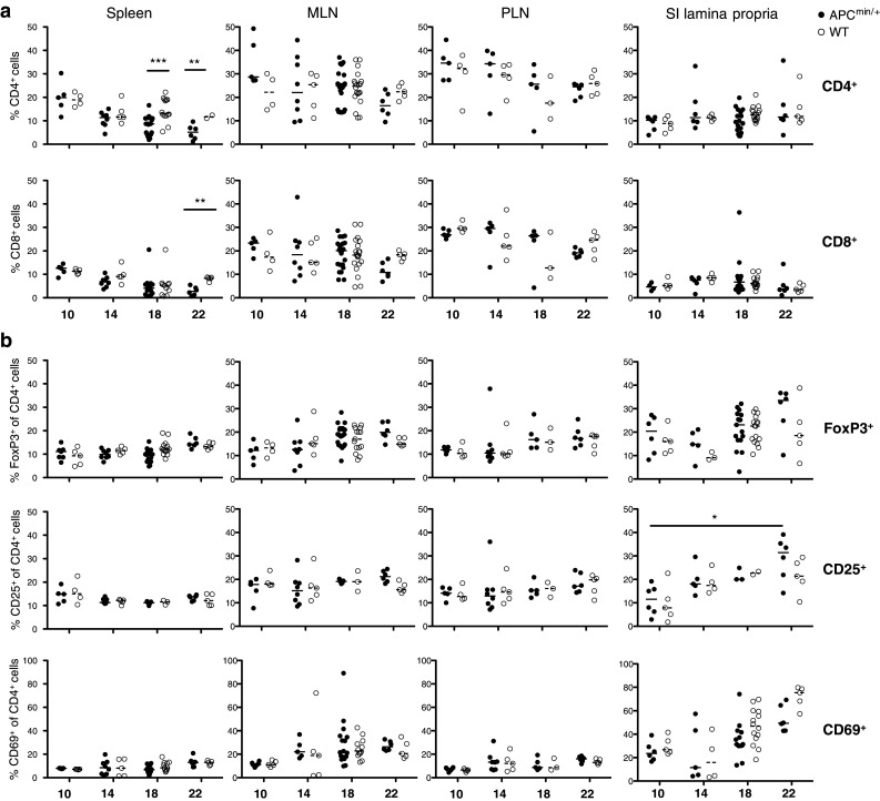
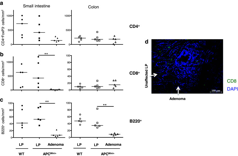
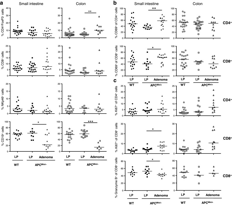
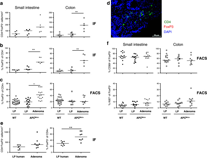
Tumor progression in the colon moves from aberrant crypt foci to adenomatous polyps to invasive carcinomas. The composition of the tumor-infiltrating leukocyte population affects the ability of the immune system to fight the tumor. T cell infiltration into colorectal adenocarcinomas, particularly T helper 1 (Th1) type T cells as well as increased regulatory T cell (Treg) frequencies, is correlated with improved prognosis. However, whether Th1 cells and Tregs are already present at the adenoma stage is not known. In this study, the APC(Min/+) mouse model of intestinal adenomatous polyposis was used to investigate tumor-associated lymphocyte subsets and the mechanisms of their accumulation into gastrointestinal adenomas. Compared to unaffected tissue, adenomas accumulated CD4(+)FoxP3(+) putative Treg in parallel with lower frequencies of conventional T cells and B cells. The accumulation of Treg was also observed in human adenomatous polyps. Despite high Treg numbers, the function of conventional T cells present in the APC(Min/+) adenomas was not different from those in the unaffected tissue. Adenomas displayed an altered chemokine balance, with higher CCL17 and lower CXCL11 and CCL25 expression than in the unaffected tissue. In parallel, CXCR3(+) Tregs were largely absent from adenomas. The data indicate that already in early stages of tumor development, the balance of lymphocyte-recruiting chemokines is altered possibly contributing to the observed shift toward higher frequencies of Treg.


Conflict of interest statement

The authors declare that they have no conflict of interest.

Figures

Fig. 1
Fig. 1
Lymphocytes frequencies in spleen, MLN, PLN and small intestine in both APCMin/+ and WT mice. The frequencies of CD8+ and CD4+ T cells (a), as well as CD4+CD69+, CD4+CD25+ and CD4+FoxP3+ (b) cells from spleen, MLN, PLN and small intestinal lamina propria were analyzed by flow cytometry at 10, 14, 18 and 22 weeks of age. Symbols represent individual mice (black APCMin/+, white WT), and lines represent median. Mann–Whitney test was used for statistical analysis *p < 0.05; **p < 0.01; ***p < 0.001
Fig. 2
Fig. 2
Lymphocyte distribution in unaffected intestinal lamina propria (LP) and adenomas. The frequencies of CD4+, CD8+ and B220+ lymphocytes were determined by immunofluorescence in small intestinal and colon sections from APCMin/+ and WT mice, and in APCMin/+ intestinal adenomas. Frequencies of conventional CD4+ T cells (a), CD8+ T cells (b) and B220+ B cells (c) are expressed as the number of cells per mm2. d Representative picture showing immunofluorescence staining of CD8+ T cells (green) in small intestinal section, displaying both adenoma and unaffected lamina propria. Symbols represent individual mice, and lines represent median. Mann–Whitney test was used for statistical analysis, **p < 0.01
Fig. 3
Fig. 3
Lymphocyte compositions in unaffected intestinal lamina propria (LP) and adenomas. Conventional, proliferating and activated T cells as well as B cells and NK cells were examined by flow cytometry in small intestine and colon. The frequencies of CD4+, CD8+, NKp46+, CD19+ (a), among all lymphocytes and CD69+ (b), Ki67+, Granzyme B+ (c), among CD4+ and CD8+ T cells were analyzed in LP of 18 weeks old APCMin/+ and WT mice, and in APCMin/+ intestinal adenomas. Symbols represent individual mice, and lines represent median. Mann–Whitney test was used as statistical analysis, *p < 0.05; **p < 0.01; ***p < 0.001
Fig. 4
Fig. 4
Regulatory T cells in murine and human unaffected intestinal tissue and adenomas. The frequencies of CD4+FoxP3+ Treg were determined by immunofluorescence (IF) and FACS in small intestinal and colon lamina propria (LP) from APCMin/+ and WT mice and in APCMin/+ adenomas, and by IF in human colon adenomatous polyps and unaffected tissue. Frequencies of CD4+FoxP3+ T cells are expressed as the number of cells per mm2 (a) or as percentage of FoxP3+ Treg among all CD4+ cells (b). c FACS analysis of CD4+FoxP3+ Treg in unaffected LP from WT mice and LP and adenoma from APCMin/+ mice. d Representative picture showing immunofluorescence staining of CD4 (green) and FoxP3 (red) in murine small intestinal adenoma section. Nuclei are stained blue with DAPI. e Frequencies of CD4+FOXP3+ T cells in human adenomatous polyps patients. f Frequencies of CD69+ and Ki67+ among CD4+FoxP3+ T cells. Symbols represent individual mice or patients, and lines represent median. Mann–Whitney test was used for statistical analysis **p < 0.01
Fig. 5
Fig. 5
Expression of chemokines and corresponding chemokine receptors by Treg in unaffected small intestine (SI) and adenomas. The expression of CCL17 (a), CCL20 (b), CCL25 (c) and CXCL11 (d) mRNA was examined in WT and APCMin/+ SI tissue and adenomas from 18-week-old mice by real-time PCR and normalized against β-actin. The frequencies of CCR4+ (a), CCR6+ (b), CCR9+ (c) and CXCR3+ (d) cells among CD4+FoxP3+ cells were analyzed by flow cytometry in WT small intestinal lamina propria (LP) and APCMin/+ LP and adenomas from 18-week-old mice. Symbols represent individual mice, and lines represent median. Mann–Whitney test was used as statistical analysis *p < 0.05; **p < 0.01
Fig. 6
Fig. 6
Cytokine producing intestinal T cells in unaffected intestinal tissue and adenomas. Lymphocytes were isolated from intestinal lamina propria (LP) from both WT and APCMin/+ mice and from adenoma tissue. The frequencies of IL-17A+ (a) and IFN-γ+ cells (b) among CD4+ or CD8+ T cells were determined by flow cytometry following PMA/ionomycin stimulation. IL-17A and IFN-γ mRNA expression (c) was determined in small intestine from both WT and APCMin/+ mice, by real-time PCR and normalized against β-actin. Symbol represents individual mice, and bar represents median

References

    1. Weitz J, Moritz K, Jurgen D, Thomas H, Galle PR, Buchler MW. Colorectal cancer. Lancet. 2005;365:153–165. doi: 10.1016/S0140-6736(05)17706-X. - DOI - PubMed
    1. Schulmann K, Reiser M, Schmiegel W. Colonic cancer and polyps. Best Pract Res Clin Gastroenterol. 2002;16:91–114. doi: 10.1053/bega.2002.0268. - DOI - PubMed
    1. Choong MK, Tsafnat G. Genetic and epigenetic biomarkers of colorectal cancer. Clin Gastroenterol Hepatol. 2012;10:9–15. doi: 10.1016/j.cgh.2011.04.020. - DOI - PubMed
    1. de la Chapelle A. Genetic predisposition to colorectal cancer. Nat Rev Cancer. 2004;4:769–780. doi: 10.1038/nrc1453. - DOI - PubMed
    1. Fridman WH, Mlecnik B, Bindea G, Pagès F, Galon J. Immunosurveillance in human non-viral cancers. Curr Opin Immunol. 2011;23:272–278. doi: 10.1016/j.coi.2010.12.011. - DOI - PubMed

Publication types