

Mucinous tumors of the ovary: current thoughts on diagnosis and management
- PMID: 24777667
- PMCID: PMC4261626
- DOI: 10.1007/s11912-014-0389-x
Mucinous tumors of the ovary: current thoughts on diagnosis and management
Abstract
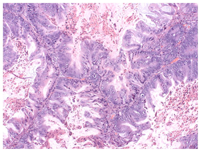
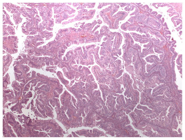
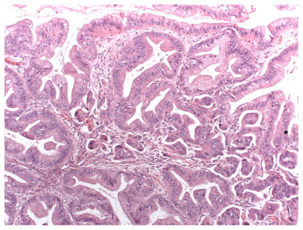
Mucinous tumors of the ovary represent a spectrum of neoplastic disorders, including benign mucinous cystadenoma, pseudomyxoma peritonei, mucinous tumors of low malignant potential (borderline), and invasive mucinous ovarian carcinoma. These tumors are related closely to each other and are distinct from other histologic subtypes of epithelial ovarian neoplasms from a clinical, histologic, and molecular standpoint. A continuum appears to be present from benign to borderline to malignant, which is different from other types of epithelial ovarian cancer. Mutational profiles are also distinct, as KRAS mutations are common, but p53 and BRCA mutations are infrequent. These characteristics lead to specific biologic behavior and guide both clinical management and research efforts in patients with mucinous ovarian tumors.
Conflict of interest statement
Figures

References
-
- Hess V, A’Hern R, Nasiri N, et al. Mucinous epithelial ovarian cancer: a separate entity requiring specific treatment. J Clin Oncol. 2004;22:1040–4. - PubMed
-
- Mondal SK, Banyopadhyay R, Nag DR, et al. Histologic pattern, bilaterality and clinical evaluation of 957 ovarian neoplasms: a 10-year study in a tertiary hospital in eastern India. J Cancer Res Ther. 2011;4:433–7. - PubMed
-
- Koonings PP. Relative frequencies of primary ovarian neoplasms: a 10-year review. Obstet Gynecol. 1989;74:921–6. - PubMed
-
- Shappell HW, Riopel MA, Smith Sehdev AE, Ronnett BM, Kurman RJ. Diagnostic criteria and behavior of ovarian seromucinous (endocervical-type mucinous and mixed cell-type) tumors: atypical proliferative (borderline) tumors, intraepithelial, microinvasive, and invasive carcinomas. Am J Surg Pathol. 2002;26:1529–41. - PubMed
Publication types
MeSH terms
Substances
Grants and funding
LinkOut - more resources
Full Text Sources
Other Literature Sources
Medical
Research Materials
Miscellaneous
