

B cell-mediated pathogenesis of ANCA-mediated vasculitis
- PMID: 24777746
- PMCID: PMC4084547
- DOI: 10.1007/s00281-014-0431-y
B cell-mediated pathogenesis of ANCA-mediated vasculitis
Abstract
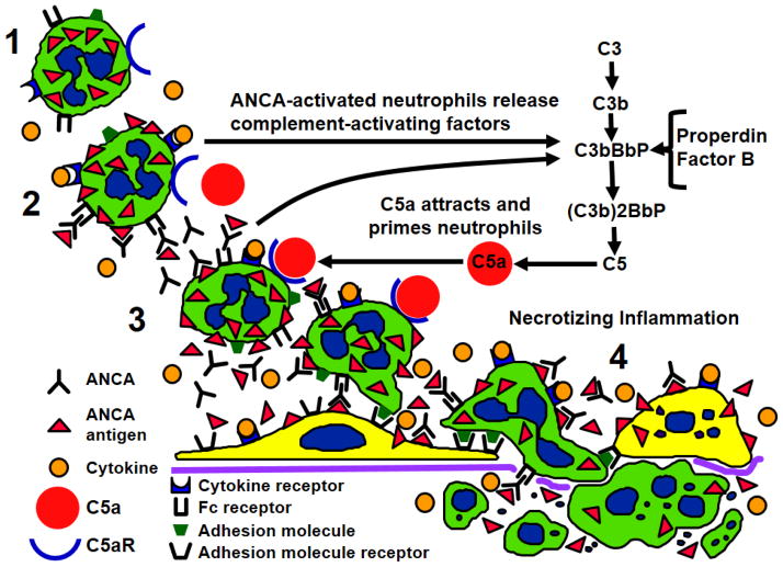
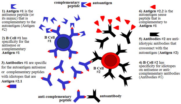
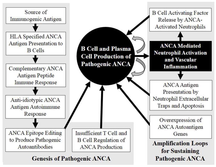
B cells and their progeny that produce and release anti-neutrophil cytoplasmic autoantibodies (ANCA) are the primary cause for an aggressive form of necrotizing small vessel vasculitis. Cytoplasmic ANCA antigens are released at the surface and in the microenvironment of cytokine-primed neutrophils. Binding of ANCA to ANCA antigens activates neutrophils by both Fc receptor engagement and direct Fab'2 binding to antigen on the cell surface. ANCA-activated neutrophils release factors that induce alternative complement pathway activation, which establishes a potent inflammatory amplification loop that causes severe necrotizing vascular inflammation. The origin of the ANCA autoimmune response is unknown but appears to involve genetically determined HLA specificities that allow the autoimmune response to develop. One putative immunogenic mechanism begins with an immune response to a peptide that is complementary to the autoantigen and evolves through an anti-idiotypic network to produce autoantibodies to the autoantigen. Another putative immunogenic mechanism begins with an immune response to a microbe-derived molecular mimic of the autoantigen resulting in antibodies that cross-react with the autoantigen. Release of neutrophil extracellular traps, apoptosis, and increased granule protein expression of ANCA antigens may facilitate the initiation of an ANCA autoimmune response, augment established pathogenic ANCA production, or both. The ANCA B cell autoimmune response is facilitated by quantitatively and qualitatively impaired T cell and B cell suppression and by release from activated neutrophils of B cell-activating factors that enhance B cell proliferation and retard B cell apoptosis.
Figures
Similar articles
-
Pathogenesis of ANCA-associated vasculitis: An update.Autoimmun Rev. 2016 Jul;15(7):704-13. doi: 10.1016/j.autrev.2016.03.007. Epub 2016 Mar 9. Autoimmun Rev. 2016. PMID: 26970490 Review.
-
Pathogenesis of vascular inflammation by anti-neutrophil cytoplasmic antibodies.J Am Soc Nephrol. 2006 May;17(5):1235-42. doi: 10.1681/ASN.2005101048. Epub 2006 Apr 19. J Am Soc Nephrol. 2006. PMID: 16624929 Review.
-
Detection by flow cytometry of anti-neutrophil cytoplasmic antibodies in a novel approach based on neutrophil extracellular traps.Autoimmunity. 2018 Sep;51(6):288-296. doi: 10.1080/08916934.2018.1527317. Autoimmunity. 2018. PMID: 30994385
-
Pathogenesis of ANCA-Associated Pulmonary Vasculitis.Semin Respir Crit Care Med. 2018 Aug;39(4):413-424. doi: 10.1055/s-0038-1673386. Epub 2018 Nov 7. Semin Respir Crit Care Med. 2018. PMID: 30404109 Free PMC article. Review.
-
Pathogenesis of ANCA-associated vasculitis.Rheum Dis Clin North Am. 2010 Aug;36(3):463-77. doi: 10.1016/j.rdc.2010.05.006. Epub 2010 Jun 23. Rheum Dis Clin North Am. 2010. PMID: 20688244 Free PMC article. Review.
Cited by
-
What is myeloperoxidase doing in ANCA-associated glomerulonephritis?Kidney Int. 2015 Nov;88(5):938-40. doi: 10.1038/ki.2015.259. Kidney Int. 2015. PMID: 26579678
-
Predictors of poor prognosis in ANCA-associated vasculitis (AAV): a single-center prospective study of inpatients in China.Clin Exp Med. 2023 Aug;23(4):1331-1343. doi: 10.1007/s10238-022-00915-z. Epub 2022 Oct 16. Clin Exp Med. 2023. PMID: 36244021 Free PMC article.
-
Comparative Histological Subtyping of Immune Cell Infiltrates in MPO-ANCA and PR3-ANCA Glomerulonephritis.Front Immunol. 2021 Oct 21;12:737708. doi: 10.3389/fimmu.2021.737708. eCollection 2021. Front Immunol. 2021. PMID: 34759920 Free PMC article.
-
Urinary cell mRNA profiling distinguishes disease activity in antineutrophil cytoplasmic antibody-associated glomerulonephritis.J Nephrol. 2023 May;36(4):1075-1077. doi: 10.1007/s40620-022-01460-4. Epub 2022 Sep 9. J Nephrol. 2023. PMID: 36083534 Free PMC article. No abstract available.
-
Chemokine expression in sera of patients with microscopic polyangiitis and granulomatosis with polyangiitis.Sci Rep. 2024 Apr 15;14(1):8680. doi: 10.1038/s41598-024-59484-8. Sci Rep. 2024. PMID: 38622321 Free PMC article.
References
-
- van der Woude FJ, Rasmussen N, Lobatto S, Wiik A, Permin H, van Es LA, van der Giessen M, van der Hem GK, The TH. Autoantibodies against neutrophils and monocytes: tool for diagnosis and marker of disease activity in Wegener’s granulomatosis. Lancet. 1985;1:425–429. - PubMed
-
- Falk RJ, Jennette JC. Anti-neutrophil cytoplasmic autoantibodies with specificity for myeloperoxidase in patients with systemic vasculitis and idiopathic necrotizing and crescentic glomerulonephritis. N Engl J Med. 1988;318:1651–1657. - PubMed
-
- Jennette JC, Falk RJ, Bacon PA, et al. Revised International Chapel Hill Consensus Conference Nomenclature of Vasculitides. Arthritis Rheum 2013. 2012;65:1–11. - PubMed
-
- Goldschmeding R, van der Schoot CE, ten Bokkel Huinink D, Hack CE, van den Ende ME, Kallenberg CGM, von dem Borne AEGK. Wegener’s granulomatosis autoantibodies identify a novel diisopropylfluorophosphate-binding protein in the lysosomes of normal human neutrophils. J Clin Invest. 1988;4:1577–1579. - PMC - PubMed
Publication types
MeSH terms
Substances
Grants and funding
LinkOut - more resources
Full Text Sources
Other Literature Sources
Research Materials
