

The Salmonella effector SteA contributes to the control of membrane dynamics of Salmonella-containing vacuoles
- PMID: 24778114
- PMCID: PMC4097631
- DOI: 10.1128/IAI.01385-13
The Salmonella effector SteA contributes to the control of membrane dynamics of Salmonella-containing vacuoles
Abstract
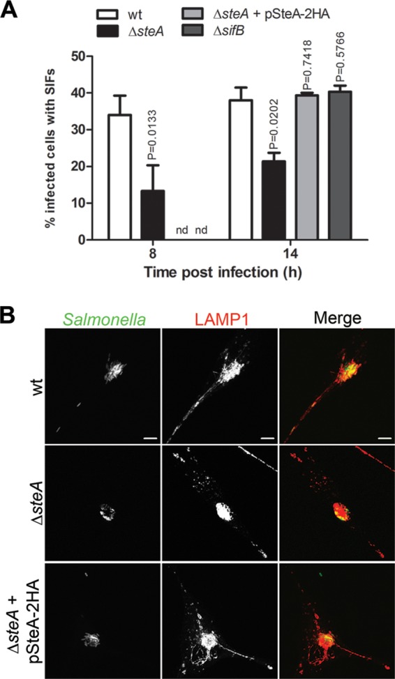
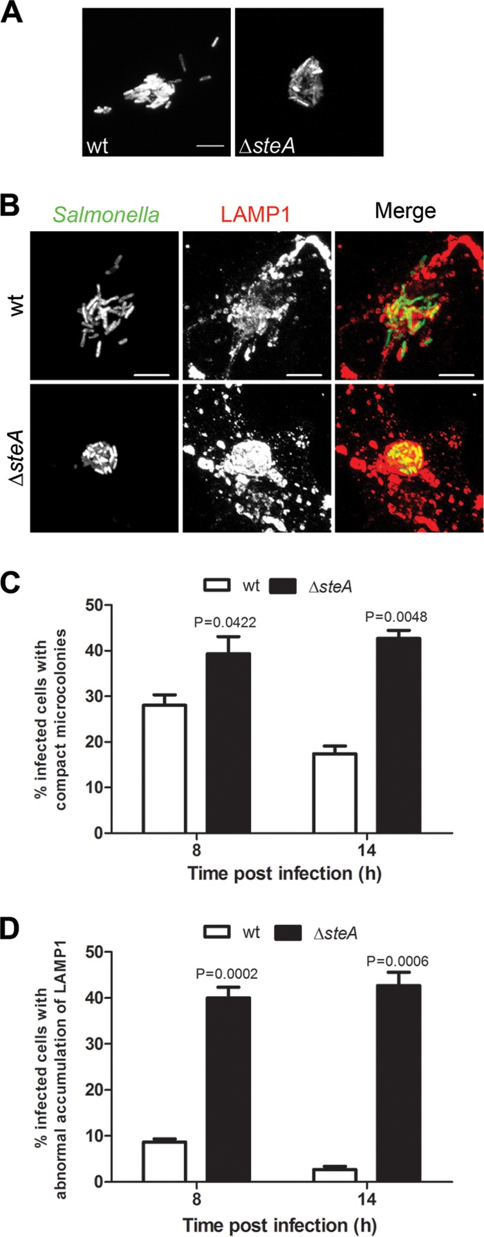
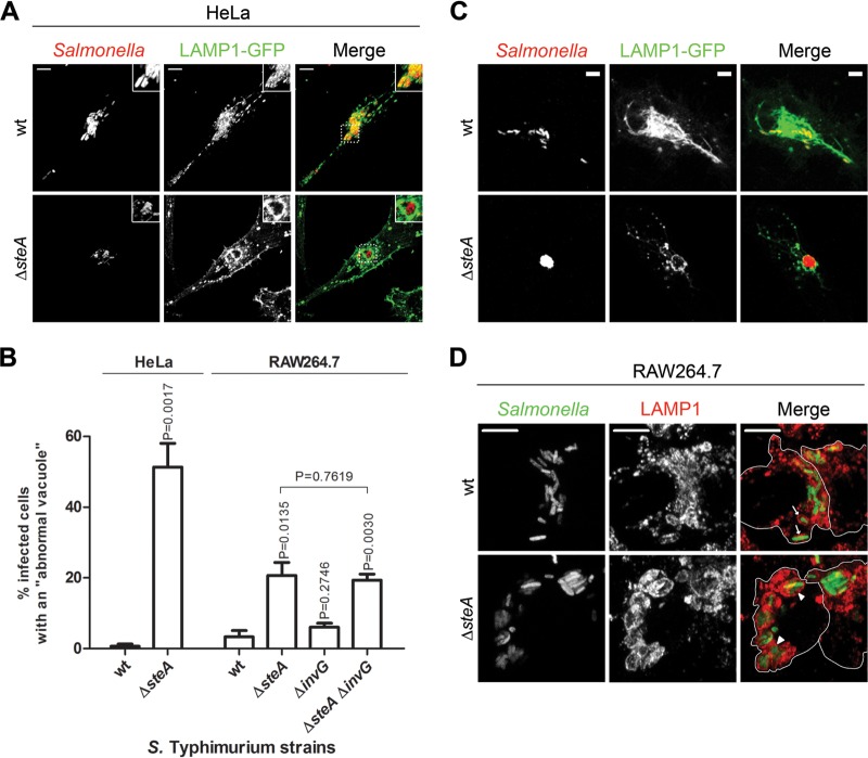
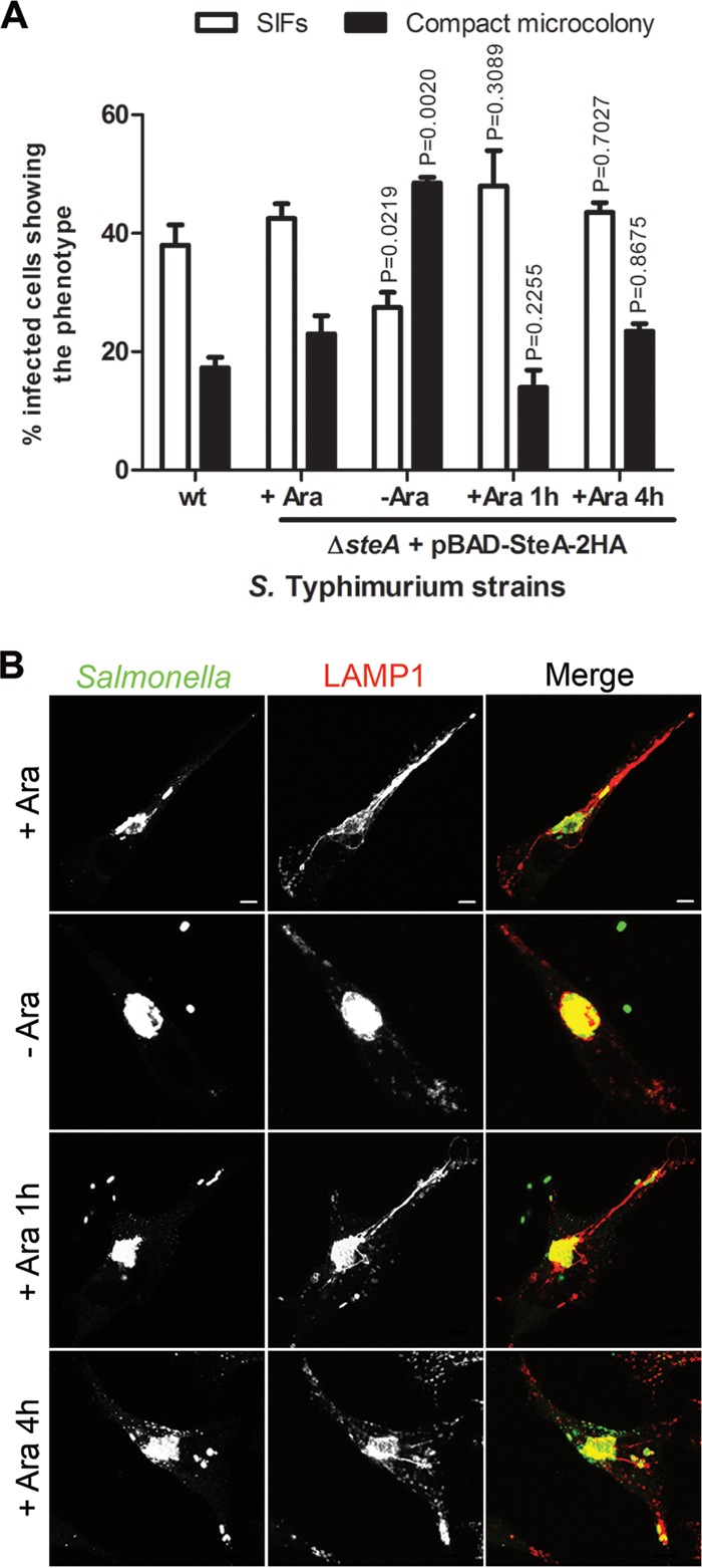
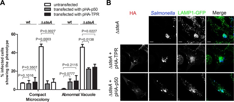
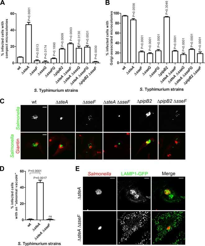
Salmonella enterica serovar Typhimurium is a bacterial pathogen causing gastroenteritis in humans and a typhoid-like systemic disease in mice. S. Typhimurium virulence is related to its capacity to multiply intracellularly within a membrane-bound compartment, the Salmonella-containing vacuole (SCV), and depends on type III secretion systems that deliver bacterial effector proteins into host cells. Here, we analyzed the cellular function of the Salmonella effector SteA. We show that, compared to cells infected by wild-type S. Typhimurium, cells infected by ΔsteA mutant bacteria displayed fewer Salmonella-induced filaments (SIFs), an increased clustering of SCVs, and morphologically abnormal vacuoles containing more than one bacterium. The increased clustering of SCVs and the appearance of vacuoles containing more than one bacterium were suppressed by inhibition of the activity of the microtubule motor dynein or kinesin-1. Clustering and positioning of SCVs are controlled by the effectors SseF and SseG, possibly by helping to maintain a balanced activity of microtubule motors on the bacterial vacuoles. Deletion of steA in S. Typhimurium ΔsseF or ΔsseG mutants revealed that SteA contributes to the characteristic scattered distribution of ΔsseF or ΔsseG mutant SCVs in infected cells. Overall, this shows that SteA participates in the control of SCV membrane dynamics. Moreover, it indicates that SteA is functionally linked to SseF and SseG and suggests that it might contribute directly or indirectly to the regulation of microtubule motors on the bacterial vacuoles.
Copyright © 2014, American Society for Microbiology. All Rights Reserved.
Figures
Similar articles
-
Salmonella Effectors SseF and SseG Interact with Mammalian Protein ACBD3 (GCP60) To Anchor Salmonella-Containing Vacuoles at the Golgi Network.mBio. 2016 Jul 12;7(4):e00474-16. doi: 10.1128/mBio.00474-16. mBio. 2016. PMID: 27406559 Free PMC article.
-
Microtubule motors control membrane dynamics of Salmonella-containing vacuoles.J Cell Sci. 2004 Mar 1;117(Pt 7):1033-45. doi: 10.1242/jcs.00949. Epub 2004 Feb 17. J Cell Sci. 2004. PMID: 14970261
-
Multiple Salmonella-pathogenicity island 2 effectors are required to facilitate bacterial establishment of its intracellular niche and virulence.PLoS One. 2020 Jun 25;15(6):e0235020. doi: 10.1371/journal.pone.0235020. eCollection 2020. PLoS One. 2020. PMID: 32584855 Free PMC article.
-
Membrane dynamics and spatial distribution of Salmonella-containing vacuoles.Trends Microbiol. 2007 Nov;15(11):516-24. doi: 10.1016/j.tim.2007.10.002. Epub 2007 Nov 5. Trends Microbiol. 2007. PMID: 17983751 Review.
-
Salmonella-containing vacuoles: directing traffic and nesting to grow.Traffic. 2008 Dec;9(12):2022-31. doi: 10.1111/j.1600-0854.2008.00827.x. Epub 2008 Oct 8. Traffic. 2008. PMID: 18778407 Review.
Cited by
-
How Bacteria Subvert Animal Cell Structure and Function.Annu Rev Cell Dev Biol. 2016 Oct 6;32:373-397. doi: 10.1146/annurev-cellbio-100814-125227. Epub 2016 May 4. Annu Rev Cell Dev Biol. 2016. PMID: 27146312 Free PMC article. Review.
-
Non-typhoidal Salmonella blood stream infection in Kuwait: Clinical and microbiological characteristics.PLoS Negl Trop Dis. 2019 Apr 15;13(4):e0007293. doi: 10.1371/journal.pntd.0007293. eCollection 2019 Apr. PLoS Negl Trop Dis. 2019. PMID: 30986214 Free PMC article.
-
Salmonella Effector SteA Suppresses Proinflammatory Responses of the Host by Interfering With IκB Degradation.Front Immunol. 2019 Dec 10;10:2822. doi: 10.3389/fimmu.2019.02822. eCollection 2019. Front Immunol. 2019. PMID: 31921113 Free PMC article.
-
Targeting of host organelles by pathogenic bacteria: a sophisticated subversion strategy.Nat Rev Microbiol. 2016 Jan;14(1):5-19. doi: 10.1038/nrmicro.2015.1. Epub 2015 Nov 23. Nat Rev Microbiol. 2016. PMID: 26594043 Review.
-
The interplay between Salmonella and host: Mechanisms and strategies for bacterial survival.Cell Insight. 2025 Feb 13;4(2):100237. doi: 10.1016/j.cellin.2025.100237. eCollection 2025 Apr. Cell Insight. 2025. PMID: 40177681 Free PMC article. Review.
References
Publication types
MeSH terms
Substances
LinkOut - more resources
Full Text Sources
Other Literature Sources
Molecular Biology Databases
Miscellaneous
