

Myeloid-derived suppressor cells in the development of lung cancer
- PMID: 24778162
- PMCID: PMC4007346
- DOI: 10.1158/2326-6066.CIR-13-0129
Myeloid-derived suppressor cells in the development of lung cancer
Abstract
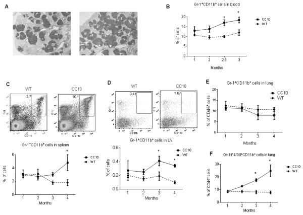
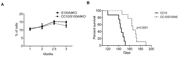
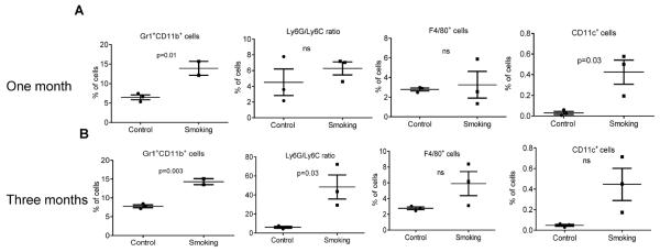
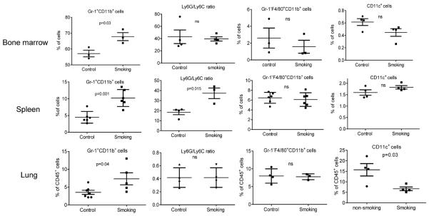
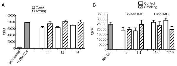
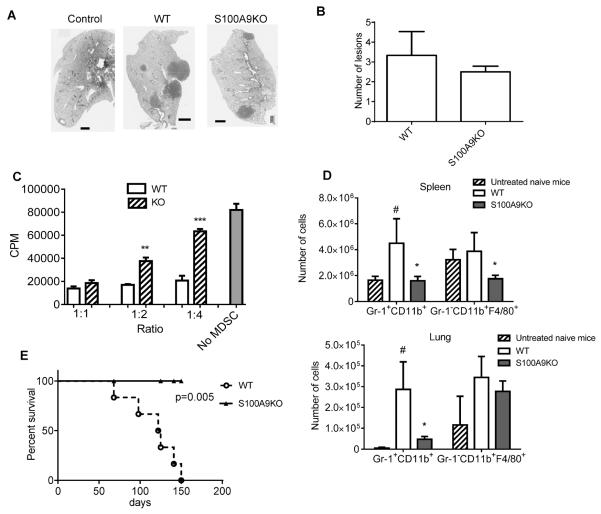
Myeloid-derived suppressor cells (MDSC) are widely implicated in immune suppression associated with tumor progression and chronic inflammation. However, very little is known about their possible role in tumor development. Here, we evaluated the role of MDSC in two experimental models of lung cancer: inflammation-associated lung cancer caused by chemical carcinogen urethane in combination with exposure to cigarette smoke; and a transgenic CC10Tg model not associated with inflammation. Exposure of mice to cigarette smoke alone resulted in significant accumulation in various organs of cells with typical MDSC phenotype (Gr-1(+)CD11b(+)). However, these cells lacked immunosuppressive activity and could not be defined as MDSC. When cigarette smoke was combined with a single dose of urethane, it led to the development of tumor lesions in lungs within 4 months. By that time, Gr-1(+)CD11b(+) cells accumulated in the spleen and lung and had potent immunosuppressive activity, and thus could be defined as MDSC. In the CC10Tg model, accumulation of immunosuppressive MDSC was observed only at 4 months of age, after the appearance of tumor lesions in the lungs. Accumulation of MDSC in both models was abrogated in S100A9 knockout mice. This resulted in a dramatic improvement in survival of mice in both models. Thus, cigarette smoke results in the expansion of immature myeloid cells lacking suppressive activity. Accumulation of bona fide MDSC in both models was observed only after the development of tumor lesions. However, MDSC played a major role in tumor progression and survival, which suggests that their targeting may provide clinical benefits in lung cancer.
©2013 AACR.
Figures
Similar articles
-
Pten null prostate epithelium promotes localized myeloid-derived suppressor cell expansion and immune suppression during tumor initiation and progression.Mol Cell Biol. 2014 Jun;34(11):2017-28. doi: 10.1128/MCB.00090-14. Epub 2014 Mar 24. Mol Cell Biol. 2014. PMID: 24662052 Free PMC article.
-
Proinflammatory S100 proteins regulate the accumulation of myeloid-derived suppressor cells.J Immunol. 2008 Oct 1;181(7):4666-75. doi: 10.4049/jimmunol.181.7.4666. J Immunol. 2008. PMID: 18802069 Free PMC article.
-
CXCL17-derived CD11b+Gr-1+ myeloid-derived suppressor cells contribute to lung metastasis of breast cancer through platelet-derived growth factor-BB.Breast Cancer Res. 2019 Feb 12;21(1):23. doi: 10.1186/s13058-019-1114-3. Breast Cancer Res. 2019. PMID: 30755260 Free PMC article.
-
The Development and Homing of Myeloid-Derived Suppressor Cells: From a Two-Stage Model to a Multistep Narrative.Front Immunol. 2020 Oct 26;11:557586. doi: 10.3389/fimmu.2020.557586. eCollection 2020. Front Immunol. 2020. PMID: 33193327 Free PMC article. Review.
-
Targeting immune suppressing myeloid-derived suppressor cells in oncology.Crit Rev Oncol Hematol. 2011 Jan;77(1):12-9. doi: 10.1016/j.critrevonc.2010.02.004. Epub 2010 Mar 20. Crit Rev Oncol Hematol. 2011. PMID: 20304669 Free PMC article. Review.
Cited by
-
Polarization of granulocytic myeloid-derived suppressor cells by hepatitis C core protein is mediated via IL-10/STAT3 signalling.J Viral Hepat. 2019 Feb;26(2):246-257. doi: 10.1111/jvh.13024. Epub 2018 Nov 19. J Viral Hepat. 2019. PMID: 30339295 Free PMC article.
-
Particulate matter air pollution as a cause of lung cancer: epidemiological and experimental evidence.Br J Cancer. 2025 Jun;132(11):986-996. doi: 10.1038/s41416-025-02999-2. Epub 2025 Apr 4. Br J Cancer. 2025. PMID: 40185876 Free PMC article. Review.
-
Bone-immune cell crosstalk: bone diseases.J Immunol Res. 2015;2015:108451. doi: 10.1155/2015/108451. Epub 2015 Apr 27. J Immunol Res. 2015. PMID: 26000310 Free PMC article. Review.
-
DEFB4A is a potential prognostic biomarker for colorectal cancer.Oncol Lett. 2020 Oct;20(4):114. doi: 10.3892/ol.2020.11975. Epub 2020 Aug 12. Oncol Lett. 2020. PMID: 32863927 Free PMC article.
-
Live or Heat-Killed Lactobacillus rhamnosus Aerosolization Decreases Adenomatous Lung Cancer Development in a Mouse Carcinogen-Induced Tumor Model.Int J Mol Sci. 2022 Oct 22;23(21):12748. doi: 10.3390/ijms232112748. Int J Mol Sci. 2022. PMID: 36361537 Free PMC article.
References
-
- Peranzoni E, Zilio S, Marigo I, Dolcetti L, Zanovello P, Mandruzzato S, et al. Myeloid-derived suppressor cell heterogeneity and subset definition. Curr Opin Immunol. 2010 - PubMed
-
- Wang L, Chang EW, Wong SC, Ong SM, Chong DQ, Ling KL. Increased myeloid-derived suppressor cells in gastric cancer correlate with cancer stage and plasma S100A8/A9 proinflammatory proteins. J Immunol. 2013;190:794–804. - PubMed
Publication types
MeSH terms
Substances
Grants and funding
LinkOut - more resources
Full Text Sources
Other Literature Sources
Medical
Research Materials
Miscellaneous
