

Physiological and structural differences in spatially distinct subpopulations of cardiac mitochondria: influence of cardiac pathologies
- PMID: 24778166
- PMCID: PMC4080170
- DOI: 10.1152/ajpheart.00747.2013
Physiological and structural differences in spatially distinct subpopulations of cardiac mitochondria: influence of cardiac pathologies
Abstract
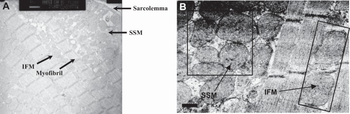
Cardiac tissue contains discrete pools of mitochondria that are characterized by their subcellular spatial arrangement. Subsarcolemmal mitochondria (SSM) exist below the cell membrane, interfibrillar mitochondria (IFM) reside in rows between the myofibrils, and perinuclear mitochondria are situated at the nuclear poles. Microstructural imaging of heart tissue coupled with the development of differential isolation techniques designed to sequentially separate spatially distinct mitochondrial subpopulations have revealed differences in morphological features including shape, absolute size, and internal cristae arrangement. These findings have been complemented by functional studies indicating differences in biochemical parameters and, potentially, functional roles for the ATP generated, based upon subcellular location. Consequently, mitochondrial subpopulations appear to be influenced differently during cardiac pathologies including ischemia/reperfusion, heart failure, aging, exercise, and diabetes mellitus. These influences may be the result of specific structural and functional disparities between mitochondrial subpopulations such that the stress elicited by a given cardiac insult differentially impacts subcellular locales and the mitochondria contained within. The goal of this review is to highlight some of the inherent structural and functional differences that exist between spatially distinct cardiac mitochondrial subpopulations as well as provide an overview of the differential impact of various cardiac pathologies on spatially distinct mitochondrial subpopulations. As an outcome, we will instill a basis for incorporating subcellular spatial location when evaluating the impact of cardiac pathologies on the mitochondrion. Incorporation of subcellular spatial location may offer the greatest potential for delineating the influence of cardiac pathology on this critical organelle.
Figures
References
-
- Asemu G, O′Connell KA, Cox JW, Dabkowski ER, Xu W, Ribeiro RF, Jr, Shekar KC, Hecker PA, Rastogi S, Sabbah HN, Hoppel CL, Stanley WC. Enhanced resistance to permeability transition in interfibrillar cardiac mitochondria in dogs: effects of aging and long-term aldosterone infusion. Am J Physiol Heart Circ Physiol 304: H514–H528, 2013 - PMC - PubMed
-
- Baseler WA, Dabkowski ER, Jagannathan R, Thapa D, Nichols CE, Shepherd DL, Croston TL, Powell M, Razunguzwa TT, Lewis SE, Schnell DM, Hollander JM. Reversal of mitochondrial proteomic loss in Type 1 diabetic heart with overexpression of phospholipid hydroperoxide glutathione peroxidase. Am J Physiol Regul Integr Comp Physiol 304: R553–R565, 2013 - PMC - PubMed
-
- Baseler WA, Dabkowski ER, Williamson CL, Croston TL, Thapa D, Powell MJ, Razunguzwa TT, Hollander JM. Proteomic alterations of distinct mitochondrial subpopulations in the type 1 diabetic heart: contribution of protein import dysfunction. Am J Physiol Regul Integr Comp Physiol 300: R186–R200, 2011 - PMC - PubMed
-
- Birkedal R, Shiels HA, Vendelin M. Three-dimensional mitochondrial arrangement in ventricular myocytes: from chaos to order. Am J Physiol Cell Physiol 291: C1148–C1158, 2006 - PubMed
Publication types
MeSH terms
Grants and funding
LinkOut - more resources
Full Text Sources
Other Literature Sources
Medical
Research Materials