. 2014 Jun 6;289(23):16409-20.
doi: 10.1074/jbc.M114.566679. Epub 2014 Apr 28.

Assembly of the respiratory mucin MUC5B: a new model for a gel-forming mucin

Affiliations

Assembly of the respiratory mucin MUC5B: a new model for a gel-forming mucin

Caroline Ridley et al. J Biol Chem. .

Abstract

Mucins are essential components in mucus gels that form protective barriers at all epithelial surfaces, but much remains unknown about their assembly, intragranular organization, and post-secretion unfurling to form mucus. MUC5B is a major polymeric mucin expressed by respiratory epithelia, and we investigated the molecular mechanisms involved during its assembly. Studies of intact polymeric MUC5B revealed a single high affinity calcium-binding site, distinct from multiple low affinity sites on each MUC5B monomer. Self-diffusion studies with intact MUC5B showed that calcium binding at the protein site catalyzed reversible cross-links between MUC5B chains to form networks. The site of cross-linking was identified in the MUC5B D3-domain as it was specifically blocked by D3 peptide antibodies. Biophysical analysis and single particle EM of recombinant MUC5B N terminus (D1D2D'D3; NT5B) and subdomains (D1, D1-D2, D2-D'-D3, and D3) generated structural models of monomers and disulfide-linked dimers and suggested that MUC5B multimerizes by disulfide linkage between D3-domains to form linear polymer chains. Moreover, these analyses revealed reversible homotypic interactions of NT5B at low pH and in high calcium, between disulfide-linked NT5B dimers, but not monomers. These results enable a model of MUC5B to be derived, which predicts mechanisms of mucin intracellular assembly and storage, which may be common to the other major gel-forming polymeric mucins.

Keywords: Analytical Ultracentrifugation; Cystic Fibrosis; Goblet Cell; Mucin; Mucus; Recombinant Protein Expression; Single Particle Analysis.


Figures

FIGURE 1.
FIGURE 1.
Calcium binding to native MUC5B. a, NaCl titration of the bound 45Ca to MUC5B by equilibrium dialysis. b, comparison of 45Ca binding to MUC5B in 10 mm (low salt; open circles) and 100 mm (high salt; filled circles) NaCl buffers. Inset: Scatchard plot of the 45Ca binding of MUC5B in 10 mm NaCl. c, More extensive analysis of 45Ca binding to MUC5B in 100 mm NaCl buffer. Inset: Scatchard plot of the 45Ca binding of MUC5B in 100 mm NaCl. B/F is the ratio of bound to free 45Ca.
FIGURE 2.
FIGURE 2.
The effect of calcium on native MUC5B self-diffusion. a, the concentration dependence of FITC-MUC5B lateral self-diffusion in the absence (open circles) or presence (filled circles) of 20 mm CaCl2. b, the lateral diffusion of microspheres (499 nm) in a range of concentrations of native MUC5B containing 0 mm (filled squares), 2 mm (open circles), or 10 mm (filled circles) CaCl2. The solid lines for 2 and 10 mm CaCl2 show the fit to the following equation (D = D0 exp(−β cυ) using least squares analysis. c, the calculated average pore size ξ determined from the tracer diffusion measurements with 499-nm microspheres over a range of concentrations of native MUC5B solution containing 2 mm (open circles) and 10 mm (filled circles) CaCl2. The ξ values were calculated from the equation ξ = (d) c−υ using the values of β and υ derived from the equation above. d, graphic of MUC5B (D-domains (white ovals), glycosylated domains (hatched boxes), Cys-domains (gray ovals), and other C-terminal domains (black circles) (8) highlighting the positions of the peptides used as immunogens for MAN5B-I, 5B-III, 5B-VI, 5B-VII, and 5B-VIII. e, Western blot analysis of N-terminal subdomain proteins (D1, D1-D2, and D3) showing the specificity of antisera that were raised against peptides from specific regions of MUC5B (MAN5B-VI, 5B-VII, 5B-VIII). f, the lateral self-diffusion of FITC-MUC5B (0.1 mg/ml) in 0 mm (black bars) or 10 mm CaCl2 (gray bars)with different MUC5B antisera or preimmune serum (PI). Error bars show the S.D. to the calculated mean for five replicates. In panels a, b, and f, measurements were performed in 0.1 m NaCl, 20 mm Tris, pH 8.0, at 25 °C, and the error bars show the S.D. to the calculated mean for five replicates.
FIGURE 3.
FIGURE 3.
Expression of recombinant MUC5B N-terminal protein domains. a, schematic showing the domains of N-terminal constructs of MUC5B. b, the expressed N-terminal construct (NT5B; D1-D2-D′-D3-domains) and subdomains (D1, D1-D2, D2-D′-D3, and D3) were analyzed by SDS-PAGE (reduced (R) and nonreduced (N)). Different NT5B protein preparations yielded varying proportions of monomer and dimer. The highlighted reduction-insensitive band detected in the D1-D2 preparation (*) was identified as desmoplakin by tandem MS. c, Western blot analysis of conditioned medium, reduced (R) and nonreduced (N), from NT5B expressed transiently in A549 cells (NT5B) and untransfected cells (−), probed with anti-His antibody. A549 cells expressed NT5B as a monomer and disulfide-linked dimer, which was comparable with NT5B expressed from 293-EBNA cells. d, SEC-MALLS analysis of NT5B showing light scattering (dashed line) and refractive index (solid line). e, molecular mass values of NT5B and expressed N-terminal MUC5B subdomains determined from SEC-MALLS analysis.
FIGURE 4.
FIGURE 4.
SEC-MALLS analysis of N-terminal subdomains of MUC5B. N-terminal subdomains of MUC5B were incubated with 5 mm CaCl2 or 5 mm EGTA, pH 7.4 or pH 6, and analyzed by SEC-MALLS. Representative graphs show the differential refractive index (RI). a–d, chromatographs for D1 (a) and D1-D2 (b) constructs showed one peak corresponding to the monomer, whereas D2-D′-D3 (c) and D3 (d) constructs showed two peaks corresponding to the dimer (first peak) and monomer (second peak). There was no effect on the chromatographic profiles of the subdomains D1, D1-D2 and D2-D′-D3 in the presence of calcium at low pH. However, D3 protein showed an effect from low pH in the presence and absence of calcium (d), indicating a potential pH-dependent aggregation.
FIGURE 5.
FIGURE 5.
Structural analysis of monomer and dimer-enriched NT5B. TEM analysis of monomer and dimer-enriched NT5B is shown. a, negatively stained particle averages for NT5B monomer (left panel) and NT5B dimer (right panel). Insets show representative class averages. b, the three-dimensional model of NT5B monomer, in three orientations. c, the three-dimensional model of NT5B dimer, in three orientations. d, SAXS analysis of the solution structure of NT5B dimer shown in three orientations. e, the monomer modeled into the dimer structure. Scale bars are 100 Å.
FIGURE 6.
FIGURE 6.
TEM analysis of dimeric D2-D′-D3 MUC5B. a, a sample field of negatively stained ”raw“ D2-D′-D3 particles. Scale bar = 100 nm. The inset shows examples of projection averages determined from the raw data. b, three-dimensional model of dimeric D2-D′-D3 in two orientations showed N-terminal dimers formed between homotypic D3-domains. Scale bar = 50 Å.
FIGURE 7.
FIGURE 7.
SAXS data for NT5B dimer. Shown are SAXS data collected for the NT5B dimer showing the scattering intensity plotted as a function of q. Inset: Guinier plot indicating Rg = 7.7 nm.
FIGURE 8.
FIGURE 8.
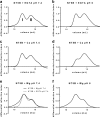
pH-dependence of calcium-mediated aggregation of the intact N-terminal domain of MUC5B. a–f, recombinant NT5B was incubated with 5 mm EGTA at pH 7.4 (a) and pH 6 (b), 5 mm CaCl2 at pH 7.4 (c) and pH 6 (d), and 5 mm MgCl2 at pH 7.4 (e) and pH 6 (f), and analyzed by SEC-MALLS. Representative graphs show the differential refractive index (n = 3). Chromatographs showed two peaks corresponding to the dimer (peak I) and monomer (peak II). The refractive index measurements showed an ∼15% reduction in the amount of NT5B protein recovered in the dimer peak in 5 mm CaCl2 at pH 6 as compared with 5 mm EGTA at pH 6. Also shown in e is the control for recombinant NT5B without MgCl2 (NT5B incubated with 5 mm EGTA at pH 7.4), which demonstrates that MgCl2 did not cause NT5B dimers to associate into higher order structures.
FIGURE 9.
FIGURE 9.
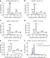
pH-dependent, calcium-mediated multimerization of disulfide-linked N-terminal MUC5B dimers. a–d, recombinant NT5B was incubated with 5 mm EGTA at pH 7.4 (a) and pH 6 (c) and 5 mm CaCl2 at pH 7.4 (b) and pH 6 (d), and analyzed by AUC to identify multimer formation (n = 3 for each combination). Representative profile showed three peaks corresponding to a monomer (peak I), a dimer (peak II), and a larger multimer (peak III). e, the calcium-mediated multimerization was studied at lower pH, and NT5B was incubated with 5 mm CaCl2 at pH 5. Percentage values of the different NT5B species were calculated from the area under the peaks. f, NT5B multimers formed at pH 6 in 5 mm CaCl2 were incubated with 10 mm EGTA at pH 7.4 overnight at 4 °C and analyzed by AUC (n = 3). The NT5B multimers were partially dissociated following incubation with EGTA at pH 7.4.
FIGURE 10.
FIGURE 10.
Model for MUC5B intracellular assembly and packaging. The MUC5B polypeptide undergoes dimerization via C-terminal to C-terminal domain (blue circles) disulfide linkage in the endoplasmic reticulum (ER). After transport to the Golgi, the dimer is glycosylated prior to assembly into linear polymers via N-terminal to N-terminal domain (red circles) disulfide links. As pH decreases and free calcium concentration increases across the secretory pathway, a conformational change occurs in the dimeric N termini, and noncovalent, calcium-mediated interactions between dimeric N termini become active. These focal links may occur within and between chains and organize MUC5B polymers for intragranular packaging. These focal links might represent the ”proteinaceous nodes“ observed in TEM images of freshly secreted MUC5B (21). Calcium binding to the charged glycans in the mucin domains (green rectangles) and its effect on mucin organization are not depicted.

Similar articles

Cited by

References

    1. Thornton D. J., Rousseau K., McGuckin M. A. (2008) Structure and function of the polymeric mucins in airways mucus. Annu. Rev. Physiol. 70, 459–486 - PubMed
    1. Hasnain S. Z., Wang H., Ghia J. E., Haq N., Deng Y., Velcich A., Grencis R. K., Thornton D. J., Khan W. I. (2010) Mucin gene deficiency in mice impairs host resistance to an enteric parasitic infection. Gastroenterology 138, 1763–1771 - PMC - PubMed
    1. Velcich A., Yang W., Heyer J., Fragale A., Nicholas C., Viani S., Kucherlapati R., Lipkin M., Yang K., Augenlicht L. (2002) Colorectal cancer in mice genetically deficient in the mucin Muc2. Science 295, 1726–1729 - PubMed
    1. Boucher R. C. (2004) New concepts of the pathogenesis of cystic fibrosis lung disease. Eur. Respir. J. 23, 146–158 - PubMed
    1. Rogers D. F. (2004) Airway mucus hypersecretion in asthma: an undervalued pathology? Curr. Opin. Pharmacol. 4, 241–250 - PubMed

Publication types

MeSH terms

LinkOut - more resources