. 2014 May 13;111(19):E2046-55.
doi: 10.1073/pnas.1405188111. Epub 2014 Apr 28.

Bacterial scaffold directs pole-specific centromere segregation

Affiliations

Bacterial scaffold directs pole-specific centromere segregation

Jerod L Ptacin et al. Proc Natl Acad Sci U S A. .

Abstract

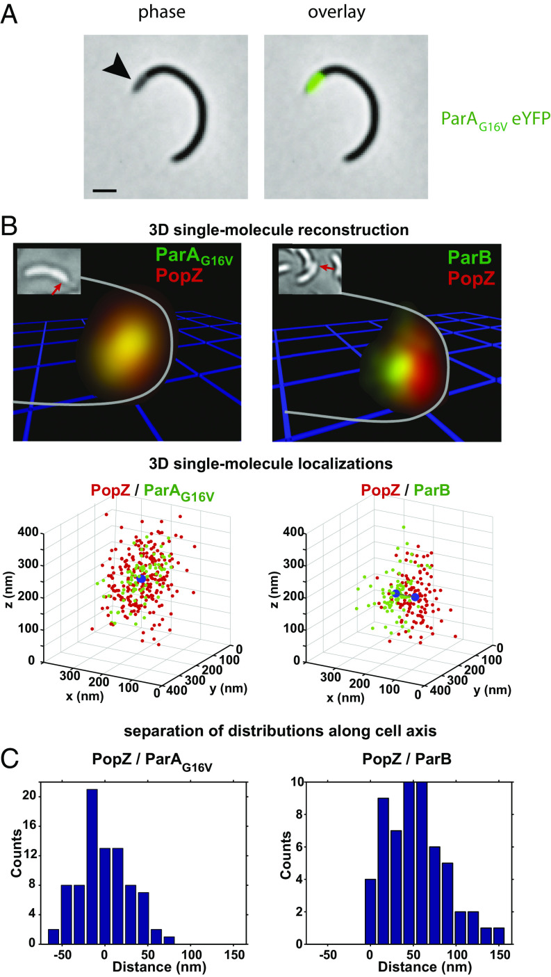
Bacteria use partitioning systems based on the ParA ATPase to actively mobilize and spatially organize molecular cargoes throughout the cytoplasm. The bacterium Caulobacter crescentus uses a ParA-based partitioning system to segregate newly replicated chromosomal centromeres to opposite cell poles. Here we demonstrate that the Caulobacter PopZ scaffold creates an organizing center at the cell pole that actively regulates polar centromere transport by the ParA partition system. As segregation proceeds, the ParB-bound centromere complex is moved by progressively disassembling ParA from a nucleoid-bound structure. Using superresolution microscopy, we show that released ParA is recruited directly to binding sites within a 3D ultrastructure composed of PopZ at the cell pole, whereas the ParB-centromere complex remains at the periphery of the PopZ structure. PopZ recruitment of ParA stimulates ParA to assemble on the nucleoid near the PopZ-proximal cell pole. We identify mutations in PopZ that allow scaffold assembly but specifically abrogate interactions with ParA and demonstrate that PopZ/ParA interactions are required for proper chromosome segregation in vivo. We propose that during segregation PopZ sequesters free ParA and induces target-proximal regeneration of ParA DNA binding activity to enforce processive and pole-directed centromere segregation, preventing segregation reversals. PopZ therefore functions as a polar hub complex at the cell pole to directly regulate the directionality and destination of transfer of the mitotic segregation machine.

Keywords: parAB; prokaryotic; replication; soj; spo0J.


Conflict of interest statement

The authors declare no conflict of interest.

Figures

Fig. 1.
Fig. 1.
PopZ interacts with ParA in vitro and in vivo. (A) PopZ is required for ParA recruitment to the Caulobacter cell pole. Images of Caulobacter cells expressing ParAG16V-eYFP protein (a monomeric form of ParA that is recruited to the cell pole) in the indicated genetic backgrounds are shown with phase-contrast and eYFP (green) fluorescence overlaid. (Scale bar, 1 µm.) (B) Purified ParA and PopZ interact directly in a concentration-dependent manner. SPR analysis using immobilized PopZ. ParA-ATP was injected at the indicated concentrations at t = 150 s, followed by buffer only (t = 300 s). Response units (R.U.s) are plotted versus time (seconds). (C) Purified ParA and PopZ interact robustly in the presence of ATP, and more weakly in the presence of ADP. SPR analysis using immobilized PopZ. ParA (1 µM) injected with ATP (green), ADP (red), no nucleotide (blue), or no ParA (black) at t = 150 s, followed by buffer only (t = 300 s). R.U.s are plotted versus time (seconds). (D) Schematic depicting a proposed ParA biochemical cycle (adapted from ref. 6). Upon ATP binding, monomeric ParA adopts a conformation that favors ParA dimerization. Dimeric ParA may interact with DNA or bind to ParB. ParB interaction stimulates ATP hydrolysis by ParA, resetting the cycle. (Left) E. coli expression/colocalization assay for mCherry-PopZ recruitment of mutant ParA-eYFP variants. Images of E. coli BL21 (DE3) cells expressing wild-type or mutant Caulobacter ParA-eYFP variant proteins (green, as indicated) in the presence of mCherry-PopZ (red) or no PopZ (right column). Colocalized red and green foci appear yellow. (Scale bar, 1 μm.)
Fig. 2.
Fig. 2.
Mutant PopZ variants are specifically defective in interacting with ParA and ParB. (A) Schematic depicting the domain structure of the PopZ protein and its functional domains R1, R2, and R3 (28). The R1 region (red) is composed of the N-terminal 24 residues, which are required for dynamic PopZ localization during the Caulobacter cell cycle and recruitment of chromosome partitioning proteins in E. coli (28, 29). The amino acid positions mutated in subsequent sections are indicated. The R2 region (white) has been shown to be a required linker between R1 and R3. The R3 region (blue) is necessary and sufficient for oligomerization (28, 29). (B) Heterologous E. coli expression/colocalization assay showing that the indicated PopZ mutant proteins [PopZ wt, PopZ-KE (E12K/R19E), PopZ-SP (S22P), and PopZ KEP (E12K/R19E/S22P)] are specifically defective in recruiting ParA or ParB in E. coli. Overlaid phase contrast and fluorescence micrographs of E. coli BL21 (DE3) cells expressing Caulobacter ParAG16V-eYFP proteins (green, middle row) and CFP-ParB (red, bottom row) in the presence of the indicated PopZ mutant protein (untagged). Foci that recruit ParA or ParB are indicated by white arrows. (Scale bar, 1 μm.) (C) PopZ variants are defective in interaction with ParA and/or ParB in vitro. SPR analysis using immobilized PopZ variants indicated. ParA-ATP (Left) or ParB (Right) at the indicated concentrations (legend) were injected at t = 150 s, followed by buffer only at t = 300 s. R.U.s are plotted versus time (seconds).
Fig. 3.
Fig. 3.
PopZ interaction with ParA is required for proper cell division and centromere segregation in Caulobacter. (A) Mutant popZ alleles that disrupt PopZ/ParA interaction cause cell morphology and centromere positioning defects in Caulobacter. Fluorescence micrographs of representative cells from strains that contain the centromere-marking cfp-parB chromosomal replacements and the indicated popZ or mcherry-popZ allele. Phase images are overlaid with CFP (green) and mCherry (red) channels (Upper) or fluorescence overlays only (Lower). Example minicells containing mCherry signal are shown for popZ-SP and popZ-KEP strains (Insets). (Scale bars, 1 μm.) (B) Mutant popZ alleles cause filamentous cell growth and cell length variability in Caulobacter. Histograms of cell lengths for the indicated strains are shown, with frequency plotted versus cell length (micrometers)(n >639 cells). (C) Mutant popZ alleles cause defective polar centromere positioning in Caulobacter. The positions of ParB foci in mixed populations of the indicated strains were quantitated and plotted versus normalized cell length (n >1,591 foci per strain). (D) Mutant popZ alleles that prevent PopZ/ParA interactions cause erratic and nonproductive ParB segregation dynamics in Caulobacter. Synchronized populations of the indicated mcherry-popZ mutant strains were subjected to time-lapse fluorescence microscopy. The positions of CFP-ParB foci along normalized cell length were determined computationally (Materials and Methods) and plotted versus time (imaging interval 5 min). ParB foci position tracks from three representative cells (red, blue, and green lines) are shown for popZ-wt, -KE, -SP, and Δ strains. Additional tracks for each strain are found in Fig. S5.
Fig. 4.
Fig. 4.
Free ParA is recruited and concentrated into a 3D PopZ matrix at the cell pole, whereas ParB is clustered on the cytoplasmic side of the PopZ complex. (A) Overexpressed PopZ forms large polar structures at the Caulobacter cell pole that recruit ParA throughout the PopZ matrix. Untagged PopZ is overexpressed in Caulobacter ΔpopZ to form extended complexes at the cell pole (black arrow indicating polar phase-bright region (18, 19) that recruit ParAG16V-eYFP (green). (Scale bar, 1 μm.) (B) Superresolution imaging reveals that ParAG16V-eYFP is recruited throughout the PopZ matrix at the cell pole, whereas ParB is clustered and offset from PopZ along the long axis of the cell. Two-color 3D superresolution reconstruction image of ParAG16V-eYFP (green) and PAmCherry-PopZ (red) (Left) and eYFP-ParB (green) and PAmCherry-PopZ (Right) localizations with respect to the estimated cell outline [white line showing the cell boundary is shown to guide the eye (Inset, viewing angle is indicated by the red arrow)]. Fluorescent molecule localizations are plotted as 3D Gaussian distributions corresponding to the localization precision of the individual emitters (Materials and Methods). Spatially overlapping red and green distributions appear yellow. Blue gridlines (500-nm squares) are included for scale and perspective. Below each reconstruction are plots of 3D localizations of ParAG16V-eYFP or eYFP-ParB (Left and Right, respectively) (green) and PAmCherry-PopZ (red) from the representative cells displayed above, showing the centroid of the distributions (blue). (C) Histograms plotting the frequency of interdistribution distances along the long cell axis for the ParA/PopZ (Left) and ParB/PopZ (Right) localization distributions obtained from 3D image cross-correlation analysis. The ParA/PopZ distributions displayed a mean difference of 0.02 ± 28.9 nm (SD, n = 83 individual cell poles), versus a mean difference of 53.1 ± 34 nm (SD, n = 57 individual cell poles) for the ParB/PopZ distributions.
Fig. 5.
Fig. 5.
ParB stimulates large-scale ParA localization into asymmetric structures near PopZ foci in E. coli. (A) ParB and PopZ direct the formation of an asymmetric ParA structure in E. coli. Images of E. coli cells expressing wild-type ParA-eYFP (green) in the presence or absence of mCherry-PopZ variants (red) and ParB expression (untagged). Fluorescence micrographs are overlaid as shown. (Scale bar, 1 μm.) Asymmetric ParA-eYFP localization (white arrowheads) occurs only when coexpressed with ParB and requires a ParA interaction-proficient PopZ variant. (B) Quantitation of mean fluorescence intensity profiles for ParA-eYFP (green) and mCherry-PopZ (red) when expressed in the presence or absence of ParB in the E. coli expression/colocalization assay. Images of representative cells were oriented with respect to the position of the polar mCherry-PopZ focus (where applicable), and the fluorescence profiles were averaged and plotted (red scale corresponds to PopZ signal (Left) and green scale corresponds to ParA signal (Right) versus normalized cell length (n >18 cells). The double hump pattern adopted by ParA-eYFP reflects accumulation on the nucleoid regions (6). Horizontal dashed lines indicate eYFP signal maxima, and vertical dashed lines indicate centers of mCherry-PopZ (red) and ParA-eYFP (green) peaks.
Fig. 6.
Fig. 6.
Model for PopZ-catalyzed ParA reassembly, a feedback mechanism to drive segregation toward the cell pole. (A) Molecular schematic for PopZ recruitment and modulation of ParA activity. A 3D matrix of PopZ (structure unknown, shown here as green lattice for clarity) recruits released/inactivated ParA molecules (purple spheres) throughout the complex. Interactions with, or increased local concentrations of, inactive ParA within the matrix facilitates localized ParA activation, and resulting activated dimeric ParA-ATP (yellow spheres) is released to encounter nearby nucleoid DNA (blue), binding with high affinity. (B) Model for PopZ modulation of ParA activity in the context of the Caulobacter cell during replication and centromere segregation. In a swarmer cell, PopZ (green) anchors ParB/parS complexes (red spheres) at the old cell pole (18, 19) and ParA-ATP (yellow spheres) localizes along the nucleoid (blue oval). Upon replication initiation, the ParB/parS complex is released from the pole (21, 31) and duplicated. Entropic forces resulting from accumulating newly replicated DNA between ParB/parS may drive centromeres apart (39), moving one ParB/parS complex away from the pole. Upon encountering the ParA/nucleoid structure, the ParB complex binds to nucleoid-bound ParA, stimulating ATP hydrolysis, releasing ParA molecules (purple spheres) from the structure and tracking along the receding edge of the shortening ParA assembly (6, 20, 21). Released ParA molecules are recruited to the cell pole by PopZ. PopZ recruitment concentrates and may allosterically stimulate ParA activation and release active molecules to bind neighboring DNA. This ParA sequestering/feedback mechanism may facilitate efficient centromere segregation and subsequent anchoring of ParB/parS to the cell pole.

Similar articles

Cited by

References

    1. Nevo-Dinur K, Govindarajan S, Amster-Choder O. Subcellular localization of RNA and proteins in prokaryotes. Trends Genet. 2012;28(7):314–322. - PubMed
    1. Lutkenhaus J. The ParA/MinD family puts things in their place. Trends Microbiol. 2012;20(9):411–418. - PMC - PubMed
    1. Leonard TA, Butler PJ, Löwe J. Bacterial chromosome segregation: Structure and DNA binding of the Soj dimer—a conserved biological switch. EMBO J. 2005;24(2):270–282. - PMC - PubMed
    1. Vecchiarelli AG, Mizuuchi K, Funnell BE. Surfing biological surfaces: Exploiting the nucleoid for partition and transport in bacteria. Mol Microbiol. 2012;86(3):513–523. - PMC - PubMed
    1. Murray H, Ferreira H, Errington J. The bacterial chromosome segregation protein Spo0J spreads along DNA from parS nucleation sites. Mol Microbiol. 2006;61(5):1352–1361. - PubMed

Publication types

MeSH terms