. 2014 May 13;111(19):6982-7.
doi: 10.1073/pnas.1402233111. Epub 2014 Apr 28.

Endophilin A1 induces different membrane shapes using a conformational switch that is regulated by phosphorylation

Affiliations

Endophilin A1 induces different membrane shapes using a conformational switch that is regulated by phosphorylation

Mark R Ambroso et al. Proc Natl Acad Sci U S A. .

Abstract

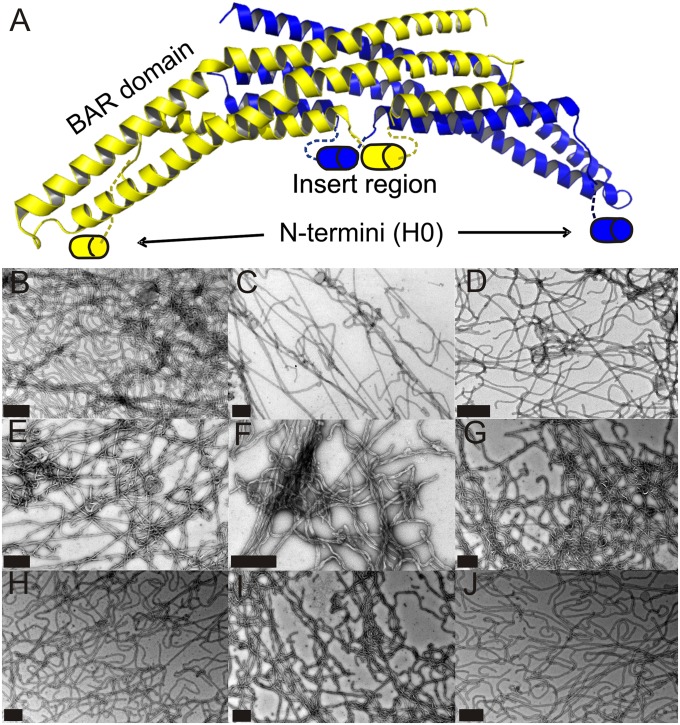
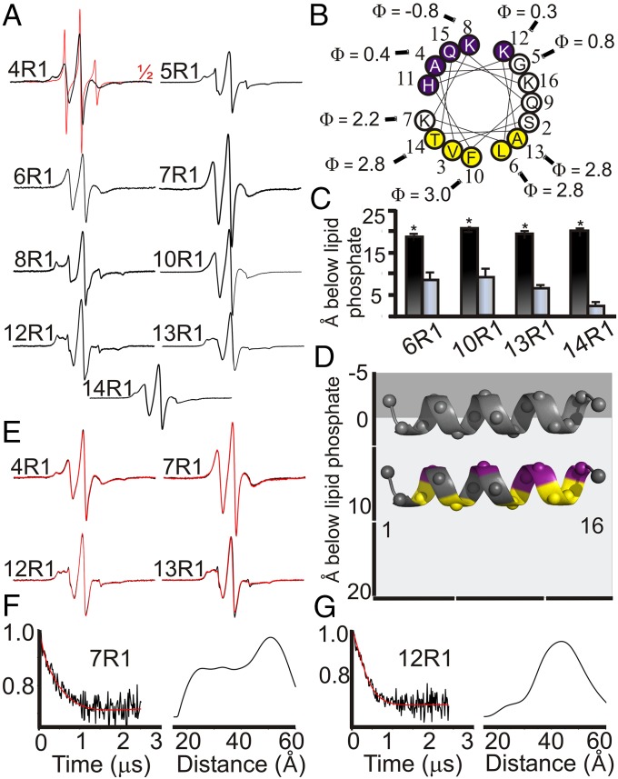
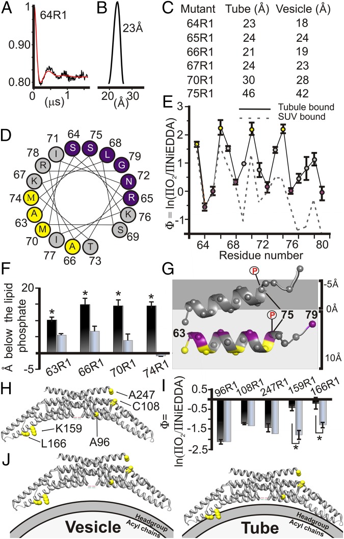
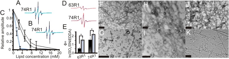
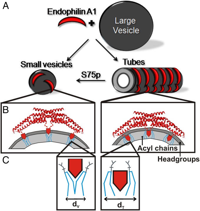
Membrane remodeling is controlled by proteins that can promote the formation of highly curved spherical or cylindrical membranes. How a protein induces these different types of membrane curvature and how cells regulate this process is still unclear. Endophilin A1 is a protein involved in generating endocytotic necks and vesicles during synaptic endocytosis and can transform large vesicles into lipid tubes or small and highly curved vesicles in vitro. By using EM and electron paramagnetic resonance of endophilin A1, we find that tubes are formed by a close interaction with endophilin A1's BIN/amphiphysin/Rvs (BAR) domain and deep insertion of its amphipathic helices. In contrast, vesicles are predominantly stabilized by the shallow insertion of the amphipathic helical wedges with the BAR domain removed from the membrane. By showing that the mechanism of membrane curvature induction is different for vesiculation and tubulation, these data also explain why previous studies arrived at different conclusions with respect to the importance of scaffolding and wedging in the membrane curvature generation of BAR proteins. The Parkinson disease-associated kinase LRRK2 phosphorylates S75 of endophilin A1, a position located in the acyl chain region on tubes and the aqueous environment on vesicles. We find that the phosphomimetic mutation S75D favors vesicle formation by inhibiting this conformational switch, acting to regulate endophilin A1-mediated curvature. As endophilin A1 is part of a protein superfamily, we expect these mechanisms and their regulation by posttranslational modifications to be a general means for controlling different types of membrane curvature in a wide range of processes in vivo.

Keywords: double electron–electron resonance; site-directed spin labeling.


Conflict of interest statement

The authors declare no conflict of interest.

Figures

Fig. 1.
Fig. 1.
Endophilin A1-induced tubulation. (A) The crystal structure of rat endophilin A1 (PDB ID code 2C08) dimer (subunits are colored yellow or blue). The N-termini (H0) and insert region are schematically illustrated as cylinders to indicate their ability to become helical upon membrane binding (17, 28, 35). Lipid tubes generated from large vesicles incubated with spin-labeled (R1) endophilin A1 derivatives are visualized by negative stain transmission EM. Representative examples are shown for N-terminal derivatives 5R1 (B), 6R1 (C), and 13R1 (D); insert region derivatives 70R1 (E), 71R1 (F), and 77R1 (G); and BAR domain derivatives 108R1 (H), 159R1 (I), and 247R1 (J). All samples were screened for thorough tubulation before further experimentation. (Scale bars: 500 nm.)
Fig. 2.
Fig. 2.
N-terminal helices penetrate deeply into the membrane. (A) CW EPR spectra of spin-labeled (R1) endophilin A1 derivatives bound to tubes (black). The EPR spectrum of position 4 in solution (red) is representative of other N-terminal sites and is shown at half amplitude. (B) Helical wheel depiction of H0 showing a hydrophilic (purple) and hydrophobic (yellow) face. Measured Φ values are shown for select residues. (C) Φ Values were converted into immersion depths for sites on the hydrophobic face of H0 on tubes (black) or in a previously elucidated vesicle-bound (gray) state (17). Depths represent the location of the nitroxide label which is typically 7–10 Å from the center of the α-helix (38). (D) A schematic model of the H0 helices on small vesicles (gray helix) or tubes (colored helix as in B) relative to the lipid headgroups (gray) and acyl chains (light gray). The helices were manually placed according to the observed average immersion depth of lipid-exposed sites, taking into account the length of the nitroxide side chain. (E) CW EPR spectra of select endophilin A1 derivatives on tubes fully labeled (black) or mixed with threefold excess of unlabeled protein (red). The overlay of the respective spectra indicates the absence of significant spin–spin interactions. (F and G) Baseline subtracted time-evolution data (black, Left) from DEER experiments of the indicated tube-bound endophilin derivatives were subjected to Tikhonov regularization (red), resulting in the shown distance distributions (Right). Error bars represent SD; n = 3 independent experiments (*P < 0.005, Student t test).
Fig. 3.
Fig. 3.
Concerted movement of BAR domain and insert region toward the membrane. (A) Baseline subtracted time-evolution data (black) from a DEER experiment of tube-bound 64R1 subjected to Tikhonov regularization (red) with (B) resulting distance distribution. (C) A comparison between intradimer distances on vesicles (28) and on tubes. (D) Local Φ maxima (yellow) and minima (purple) fall onto a hydrophobic or hydrophilic face of a helical wheel. (E) Φ as function of labeling position on tubes (solid, colored as in D) and on vesicles (28) (dashed). (F) Immersion depth after calibration for sites on the hydrophobic face of the insert region on vesicles (28) (gray) and on tubes (black). (G) A schematic model of the insert region on vesicles (gray helix) and tubes (colored as in D) relative to the lipid headgroups (dark gray, negative depth values) and the acyl chains (light gray, positive depth values). The helices were manually placed as described in Fig. 2. The phosphorylation site S75 moves from the acyl chain environment to the aqueous environment (illustrated phosphate groups). (H) Crystal structure of rat endophilin A1 dimer (PDB ID code 2C08) showing the locations of spin-labeled sites. (I) Bar graph comparing Φ values measured for sites on the concave and convex surfaces of the BAR domain when bound to tubes (black) or vesicles (gray). (J) Schematic illustration of the location of the BAR domain relative to the bilayer when bound to vesicles (Left) or tubes (Right). Error bars represent SD; n represents at least three independent experiments (*P < 0.01, Student t test).
Fig. 4.
Fig. 4.
Phosphomimetic S75D mutation destabilizes tubes by reducing membrane immersion depth of the insert region. CW EPR spectra of S75D-74R1 incubated with 0 mM (black), 2.5 mM (purple), and 10 mM (teal) of (A) vesicles composed of a 5:2:1:1 molar ratio of l-α-phosphatidylcholine, l-α-phosphatidylethanolamine, l-α-phosphatidylserine, and cholesterol (4) or (B) vesicles composed of 2-Dioleoyl-sn-Glycero-3-[Phospho-rac-(1-glycerol)] sodium salt and 1,2-Dioleoyl-sn-glycero-3-phosphoethanolamine (2:1). (C) Spectral amplitudes for S75-74R1 (solid lines) and S75D-74R1 (dashed lines) from the experiments in A (black lines) or B (blue lines) are plotted as function of lipid concentration. (D) CW EPR spectra of tube-bound endophilin A1 labeled at positions 63 or 74 with (black) or without (red) the S75D mutation. All spectra are normalized to the same number of spins. (E) Φ Values of 63R1 and 74R1 with (black) and without (gray) the S75D mutation incubated with 2-Dioleoyl-sn-Glycero-3-[Phospho-rac-(1-glycerol)] sodium salt and 1,2-Dioleoyl-sn-glycero-3-phosphoethanolamine (2:1). Negative stain EM shows S75-63R1 forming stable tubes after 24 h (F), whereas S75D-63R1 produces mainly vesicular structures (G). Similar results were obtained for S75-74R1 (H) and S75D-74R1 (I) as well as S75 (J) and S75D (K) in a WT background. (Scale bars: 0.5 μm.) Error bars represent SD; n represents at least three independent experiments (*P < 0.005, Student t test).
Fig. 5.
Fig. 5.
Schematic illustration of endophilin A1 tube and vesicle binding and its modulation by phosphorylation. (A) The incubation of endophilin A1 and large lipid vesicles can result in vesiculation or tubulation. (B) On small vesicles (Left), endophilin A1 predominantly uses its amphipathic helices (red pentagons) rather than the BAR domain for membrane binding. The location of the helices is optimized for stabilizing membrane curvature by wedging into the headgroup region (dark gray) and thereby generating a splitting force between neighboring lipids (C, blue lines). Endophilin A1 binds tubes (A, Right) in a highly oligomeric, anisotropic manner (34, 35), and moves its amphipathic helices deeper into the acyl chains (B, Right), filling more space within the acyl chain region (light gray), which more optimally pushes entire lipids apart (C, Right). The difference in lipid area the helices take up in the membrane is greater for tubes (dt) than for vesicles (dv). Simultaneously, the BAR domain moves into contact with the lipid headgroups (B, Right). For simplicity only the outer leaflet of the membrane is shown in B. LRRK2 phosphorylation introduces a negative charge at S75, a site submerged in the acyl chain region of tubes. This modification destabilizes tubes and favors vesicles as illustrated in A.

References

    1. Rao Y, Haucke V. Membrane shaping by the Bin/amphiphysin/Rvs (BAR) domain protein superfamily. Cell Mol Life Sci. 2011;68(24):3983–3993. - PMC - PubMed
    1. McMahon HT, Gallop JL. Membrane curvature and mechanisms of dynamic cell membrane remodelling. Nature. 2005;438(7068):590–596. - PubMed
    1. Qualmann B, Koch D, Kessels MM. Let’s go bananas: Revisiting the endocytic BAR code. EMBO J. 2011;30(17):3501–3515. - PMC - PubMed
    1. Matta S, et al. LRRK2 controls an EndoA phosphorylation cycle in synaptic endocytosis. Neuron. 2012;75(6):1008–1021. - PubMed
    1. Bergmann C, et al. Oligophrenin 1 (OPHN1) gene mutation causes syndromic X-linked mental retardation with epilepsy, rostral ventricular enlargement and cerebellar hypoplasia. Brain. 2003;126(Pt 7):1537–1544. - PubMed

Publication types