. 2014 May 13;111(19):6940-5.
doi: 10.1073/pnas.1324050111. Epub 2014 Apr 28.

Large, stratified, and mechanically functional human cartilage grown in vitro by mesenchymal condensation

Affiliations

Large, stratified, and mechanically functional human cartilage grown in vitro by mesenchymal condensation

Sarindr Bhumiratana et al. Proc Natl Acad Sci U S A. .

Abstract

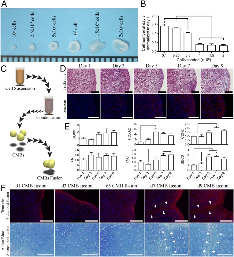
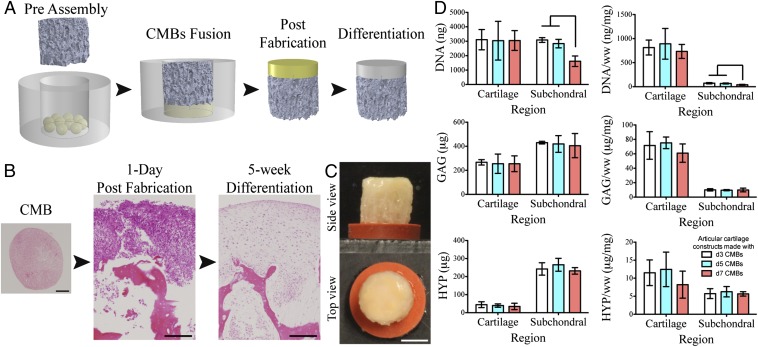
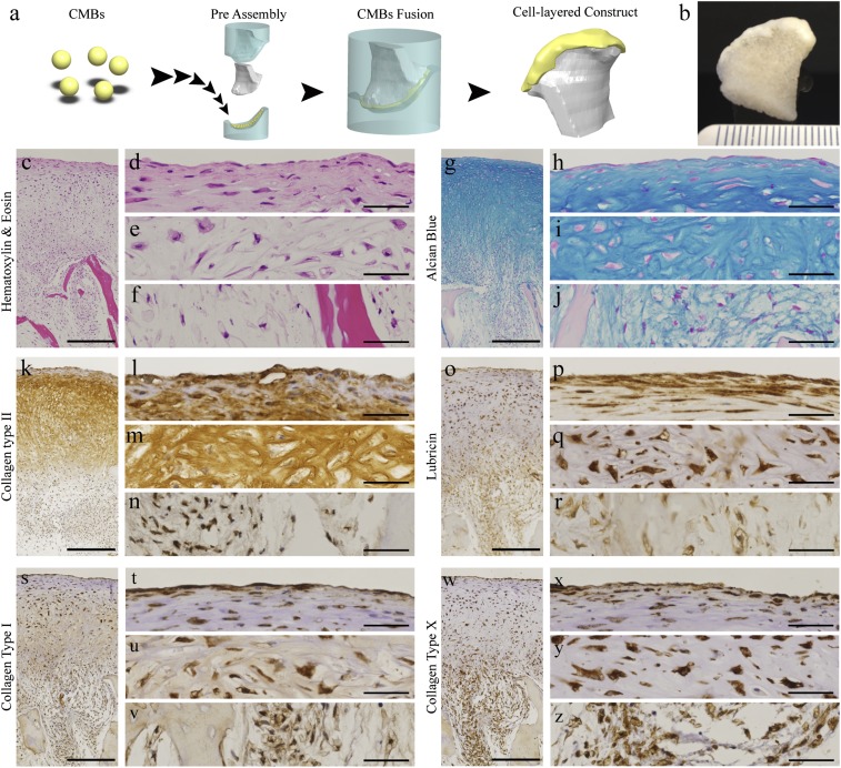
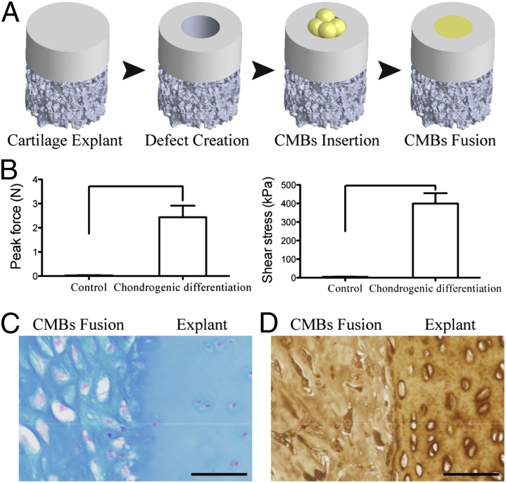
The efforts to grow mechanically functional cartilage from human mesenchymal stem cells have not been successful. We report that clinically sized pieces of human cartilage with physiologic stratification and biomechanics can be grown in vitro by recapitulating some aspects of the developmental process of mesenchymal condensation. By exposure to transforming growth factor-β, mesenchymal stem cells were induced to condense into cellular bodies, undergo chondrogenic differentiation, and form cartilagenous tissue, in a process designed to mimic mesenchymal condensation leading into chondrogenesis. We discovered that the condensed mesenchymal cell bodies (CMBs) formed in vitro set an outer boundary after 5 d of culture, as indicated by the expression of mesenchymal condensation genes and deposition of tenascin. Before setting of boundaries, the CMBs could be fused into homogenous cellular aggregates giving rise to well-differentiated and mechanically functional cartilage. We used the mesenchymal condensation and fusion of CMBs to grow centimeter-sized, anatomically shaped pieces of human articular cartilage over 5 wk of culture. For the first time to our knowledge biomechanical properties of cartilage derived from human mesenchymal cells were comparable to native cartilage, with the Young's modulus of >800 kPa and equilibrium friction coeffcient of <0.3. We also demonstrate that CMBs have capability to form mechanically strong cartilage-cartilage interface in an in vitro cartilage defect model. The CMBs, which acted as "lego-like" blocks of neocartilage, were capable of assembling into human cartilage with physiologic-like structure and mechanical properties.

Keywords: biomimetic; cartilage mechanics; cartilage repair; regenerative medicine; tissue engineering.


Conflict of interest statement

The authors declare no conflict of interest.

Figures

Fig. 1.
Fig. 1.
Generation and fusion of CMBs formed in vitro. (A) Day-3 CMBs created from 1 × 10⁵ to 2 × 10⁶ hMSCs. (B) The DNA content in the condensed cellular body was quantified and normalized to the initial seeding DNA content. (C) Schematic of the condensation and fusion of CMBs. (D) At day 7, CMBs began to develop tangential cellular lining along the outer surface (Upper Row, Trichrome). The periphery surface of d7 and d9 CMBs stained for tenascin, an indication of boundary setting. (E) Boundary of CMBs was also characterized by high expression of TNC and SDC3 genes, and increased expression of HOXA2 and CDH2 genes. SOX9, chondrogenesis factor, gradually increases as CMBs became more mature. Fibronectin expression, FN, remained constant. (F) Fusion of CMBs at different developmental stages. At days 7–9 postfusion, all CMBs showed tenascin deposition (Upper Row) at the periphery, in contrast to early stage CMBs (d1-5). Chondrogenesis in fused early stage CMBs resulted in a homogenous glycosaminoglycan structure (Lower Row), whereas the border between the adjacent CMBs was clearly seen in fused late stage CMBs (arrow). [Scale bars: (A) 1 mm; (D) 100 μm; and (F) 200 μm.] The lines in B and E indicate significant differences between the groups (P < 0.05).
Fig. 2.
Fig. 2.
Effect of the CMB developmental stage on cartilage formation. (A) To form articular cartilage on bone substrate, CMBs were placed into a PDMS ring, a bone scaffold was inserted and pressed onto CMBs to cause CMBs to fuse and penetrate inside the scaffold pores resulting in a composite osteochondral construct. After differentiation, the cellular layer formed into cartilage and integrated with the porous scaffold. (B) CMBs and osteochondral constructs at d1 and week 5 post fusion (H&E). Histological and immunohistochemical sections of the bioengineered cartilage and subchondral bone indicating appropriate matrix composition and cartilage formation. (C) Top and side views of the articular cartilage plug with the fused CMBs developed into thick cartilage layer covering the whole construct surface. (D) After 5 wk of chondrogenic induction, cartilage layers created from d3, d5, and d7 CMBs had similar contents of DNA, glycosaminoglycan (GAG), and hydroxyproline (HYP). Subchondral regions had similar GAG and HYP contents, and significantly lower DNA content for d7 CMBs, suggesting reduced migratory and integrative ability. [Scale bars: (B) 200 μm and (C) 2 mm.] Lines in D indicate significant differences between the groups (P < 0.05).
Fig. 3.
Fig. 3.
The utility of CMB fusion for engineering large, anatomically shaped cartilage (A) CMBs were placed on the cartilage side of a mold in the exact shape of the condyle, an anatomically shaped porous scaffold was placed on the other side, and the two-piece mold was press-fit. CMBs fused together and adhered to the scaffold as a thick cellular layer along the articular surface. (B) Anatomical layer of articular cartilage on underlying bone after 5 wk of cultivation. (CZ) Histological and immunohistochemical analysis of cartilage matrix stained for (CF) H&E, (GJ) Alcian blue for GAG, (KN) anticollagen type II, (OR) antilubricin, (SV) anticollagen type I, and (WZ) anticollagen type X. (C, G, K, O, S, and W) Low-magnification images. (D–F, H–J, L–N, P–R, T–V, and X–Z) High magnification images. (Scale bar: 500 μm in low-magnification images and 50 µm in high-magnification images.)
Fig. 4.
Fig. 4.
Utility of CMBs for cartilage repair. (A) Cartilage defects (1.5 mm diameter) were created by biopsy punch and filled with pressed CMBs to fuse and fill the defect. (B) After 5 wk of chondrogenic induction, CMBs formed cartilage tissue that integrated with the native cartilage, as evidenced by the measured peak force and shear stress needed to break the integration surface. The high integration strength was due to the structural integration of glycosaminoglycan and collagen type II as indicated by (C) Alcian blue and (D) anticollagen type II immunohistochemistry stain. Lines in B indicate significant differences between the groups (P < 0.05). (Scale bar: 50 μm.)

Comment in

References

    1. Langer RS, Vacanti JP. Tissue engineering: The challenges ahead. Sci Am. 1999;280(4):86–89. - PubMed
    1. Nakao K, et al. The development of a bioengineered organ germ method. Nat Methods. 2007;4(3):227–230. - PubMed
    1. Tew SR, Kwan AP, Hann A, Thomson BM, Archer CW. The reactions of articular cartilage to experimental wounding: Role of apoptosis. Arthritis Rheum. 2000;43(1):215–225. - PubMed
    1. Quinn TM, Allen RG, Schalet BJ, Perumbuli P, Hunziker EB. Matrix and cell injury due to sub-impact loading of adult bovine articular cartilage explants: Effects of strain rate and peak stress. J Orthop Res. 2001;19(2):242–249. - PubMed
    1. Chen CT, Burton-Wurster N, Lust G, Bank RA, Tekoppele JM. Compositional and metabolic changes in damaged cartilage are peak-stress, stress-rate, and loading-duration dependent. J Orthop Res. 1999;17(6):870–879. - PubMed

Publication types

MeSH terms