. 2014 May 13;111(19):7132-7.
doi: 10.1073/pnas.1403285111. Epub 2014 Apr 28.

Neurosteroids promote phosphorylation and membrane insertion of extrasynaptic GABAA receptors

Affiliations

Neurosteroids promote phosphorylation and membrane insertion of extrasynaptic GABAA receptors

Armen M Abramian et al. Proc Natl Acad Sci U S A. .

Abstract

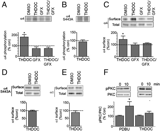
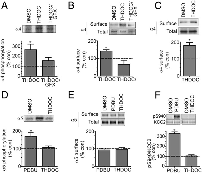
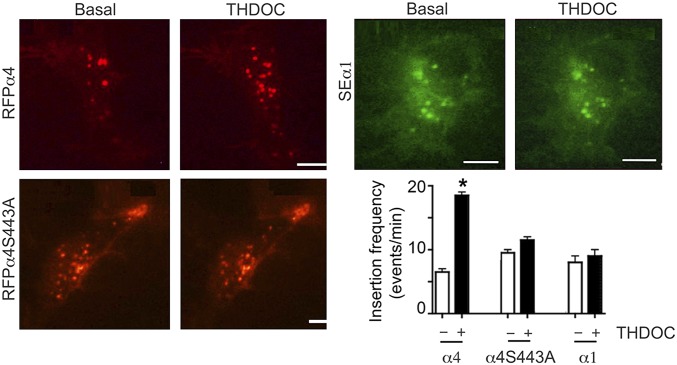
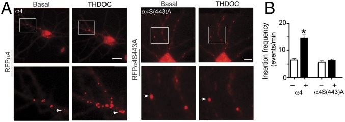
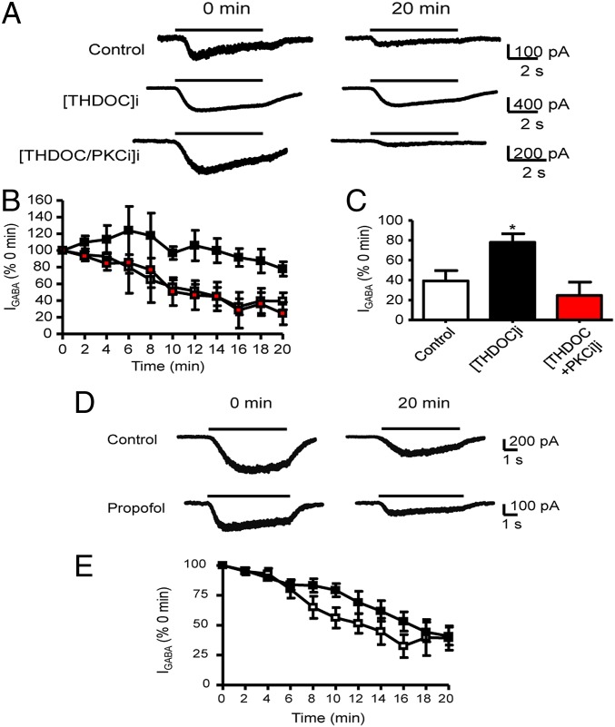
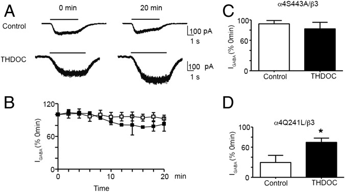
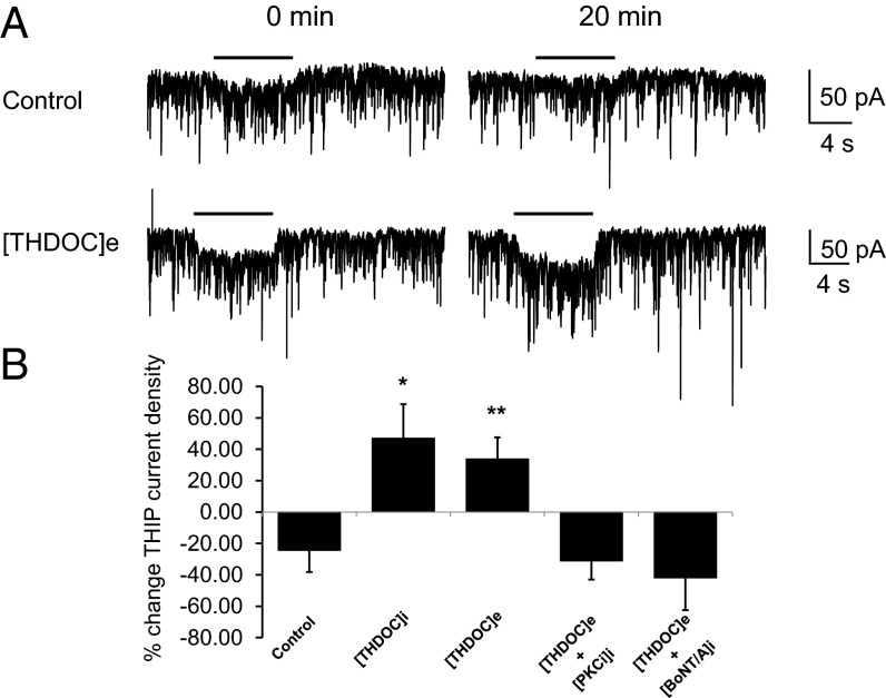
Neurosteroids are synthesized within the brain and act as endogenous anxiolytic, anticonvulsant, hypnotic, and sedative agents, actions that are principally mediated via their ability to potentiate phasic and tonic inhibitory neurotransmission mediated by γ-aminobutyric acid type A receptors (GABAARs). Although neurosteroids are accepted allosteric modulators of GABAARs, here we reveal they exert sustained effects on GABAergic inhibition by selectively enhancing the trafficking of GABAARs that mediate tonic inhibition. We demonstrate that neurosteroids potentiate the protein kinase C-dependent phosphorylation of S443 within α4 subunits, a component of GABAAR subtypes that mediate tonic inhibition in many brain regions. This process enhances insertion of α4 subunit-containing GABAAR subtypes into the membrane, resulting in a selective and sustained elevation in the efficacy of tonic inhibition. Therefore, the ability of neurosteroids to modulate the phosphorylation and membrane insertion of α4 subunit-containing GABAARs may underlie the profound effects these endogenous signaling molecules have on neuronal excitability and behavior.

Keywords: PKC; current rundown; receptor insertion; tonic current.


Conflict of interest statement

The authors declare no conflict of interest.

Figures

Fig. 1.
Fig. 1.
Neurosteroids regulate the phosphorylation and cell surface expression of recombinant GABAARs containing α4 subunits. (A) HEK cells expressing α4β3 receptors were labeled with 1 mCi/mL 32P-orthosphosphoric acid and treated for 10 min with DMSO (control), 100 nM THDOC, or 20 μM GFX/100 nM THDOC. Phosphorylation of α4 was measured using immunoprecipitation with subunit-specific antibodies and data normalized to vehicle-treated samples. (B) The effects of 100 nM THDOC on the phosphorylation of receptors composed of α4S443A subunits were measured as outlined above. (C) Cells expressing α4β3 subunits were treated for 10 min with DMSO (control), 100 nM THDOC, or 20 μM GFX/100 nM THDOC and labeled with NHS-biotin. The resulting cell surface and total fractions were then immunoblotted with α4 subunit antibodies. The ratio of cell surface to total α4 subunit immunoreactivity was determined and normalized to vehicle-treated control (dotted line; 100%). (D) The effects of 100 nM THDOC on the cell surface accumulation of receptors composed of α4(S443A) and β3 subunits were measured as outlined above. (E) The effects of 100 nM THDOC on the cell surface accumulation of receptors composed of α1 and β3 subunits were measured as outlined above. (F) HEK cells were treated with 100 nM PDBU or 100 nM THDOC for 10 min and then immunoblotted with pT638 and a PKC antibody that recognizes the α, βI–II, and γ subtypes of PKC. The ratio of pT638/PKC immunorecativity was determined and normalized to levels seen at t = 0. *, significantly different to control in all panels (P < 0.05; n = 4–6).
Fig. 2.
Fig. 2.
Neurosteroids selectively regulate the phosphorylation and cell surface expression of GABAARs containing α4 subunits in hippocampal slices. (A) We labeled 350 μm hippocampal slices from 8- to 12-wk-old mice with 1 mCi/mL 32P-orthosphosphoric acid and treated them for 10 min with DMSO (control), 100 nM THDOC, or 20 μM GFX/100 nM THDOC. Phosphorylation of α4 was measured using immunoprecipitation with subunit-specific antibodies and data normalized to vehicle-treated samples (dotted line; 100%). (B) Hippocampal slices were treated as above and subject to biotinylation. Cell surface and total fractions were then immunoblotted with α4 subunit antibodies. The ratio of cell surface to total α4 subunit immunoreactivity was determined and normalized to vehicle-treated controls. (C) Cell surface expression levels of the α4 subunit were determined in hippocampal slices from C57/Bl6 δ-KO mice treated for 10 min with DMSO (control) or 100 nM THDOC as detailed above. (D) Phosphorylation of the α5 subunit was measured in 32P-labeled hippocampal slices using immunoprecipitation with subunit-specific antibodies and data normalized to vehicle-treated samples. (E) Hippocampal slices were treated as above and subject to biotinylation. Cell surface and total fractions were then immunoblotted with α5 subunit antibodies. The ratio of cell surface to total α5 subunit immunoreactivity was determined and normalized to vehicle-treated controls. (F) Hippocampal slices were treated with the respective agents and then immunoblotted with pS940 and KCC2 antibodies. The ratio of pS940/KCC2 immunoreactivity was determined and normalized to vehicle-treated controls (dotted line; 100%).
Fig. 3.
Fig. 3.
Neurosteroids modulate the membrane insertion of GABAARs dependent upon S443 in the α4 subunit. HEK cells expressing RFPα4β3, RFPα4(S443A)β3, or SEα1β3 receptors were imaged by TIRF for 5 min before (basal) and after 20-min incubation with 100 nM THDOC. These data were then used to determine the insertion frequency for each α subunit construct in the absence and presence of THDOC, as shown in the lower right panel. *, significantly different from control (P < 0.05; n = 5).
Fig. 4.
Fig. 4.
Neurosteroids modulate the membrane insertion of the α4 subunit in hippocampal neurons. (A) The 10–15 Div hippocampal neurons expressing RFPα4 or RFPα4(S443A) subunits were subject to TIRF for 5 min before (basal) and 5 min after 20-min incubation at 37 °C with 100 nM THDOC. The images in the lower panels are enlargements of the boxed regions in the upper panels. (B) The total number of insertion events per minute was then calculated in the absence and presence of THDOC. *, significantly different from control (t test, P < 0.05; n = 5–7).
Fig. 5.
Fig. 5.
Internal THDOC prevents GABAA α4β3 receptor-mediated current rundown via a PKC-dependent process. (A) The 1 μM (∼EC50) GABA-activated currents (IGABA) recorded at 0 and 20 min after the start of the experiment (defined as t = 0 min and 100%). Whole-cell currents were recorded from HEK cells expressing α4β3 receptors in the presence of internally applied vehicle (DMSO) control (upper currents), internal 100 nM THDOC ([THDOC]i; middle currents), or internal THDOC plus 200 nM PKC19–36 inhibitor peptide ([THDOC/PKCi]i; lower currents). The black line above the current traces represents the application of GABA. (B) Time dependence relationship for (IGABA) recorded in the presence of either internally applied vehicle control (DMSO) (white square), 100 nM THDOC (black square), or 100 nM THDOC and 200 nM PKC19–36 inhibitor peptide (red square). *, significantly different from control DMSO and PKC inhibitor peptide (P = 0.025, n = 3–6). (C) Bar graph of the (IGABA) at t = 20 min compared with currents at t = 0 min for α4β3 receptors in control conditions (white bar) or in the presence of internal 100 nM THDOC (black bar) or internal THDOC plus PKC19–36 inhibitor peptide (red bar). (D) GABA-activated currents recorded from α4β3 receptors at 0 and 20 min after the start of the experiment either in the absence (control, upper currents) or presence of internal 3 μM propofol (lower currents). (E) The time dependence relationship for (IGABA) recorded in the presence of either internally applied vehicle control (white square) or Propofol (black square).
Fig. 6.
Fig. 6.
Prevention of α4β3 receptor-mediated current rundown by THDOC is dependent upon S443 in the α4 subunit but independent of THDOC-mediated allosteric modulation. (A) (IGABA) recorded at 0 and 20 min after the start of the experiment. Whole-cell currents were recorded from HEK293 cells expressing α4(S443A)β3 receptors in the presence of internally applied vehicle control (upper currents) or internal 100 nM THDOC ([THDOC]i; lower currents). The black line above the current traces represents the application of GABA. (B) Time dependence relationship for (IGABA) recorded in the presence of either internally applied vehicle control (white square) or 100 nM THDOC (black square). (C) Bar graph of the relative (IGABA) at t = 20 min compared with current at t = 0 min for α4(S443A)β3 receptors in control conditions (white bar) or perfused internally with 100 nM THDOC (black bar). (D) Bar graph of the relative (IGABA) at t = 20 min compared with current at t = 0 min for α4(Q241L)β3 receptors in control conditions (white bar) or perfused internally with 100 nM THDOC (black bar). *, significantly different from control (t test, P = 0.026; n = 3–6).
Fig. 7.
Fig. 7.
THDOC selectively enhances tonic current in hippocampal neurons. (A) THIP-activated currents recorded at 0 and 20 min after the start of the experiment. Whole-cell currents were recorded from 21–29 Div rat hippocampal neurons in the presence of internally applied vehicle control (upper currents) or following a 10-min exposure to extracellular 100 nM THDOC ([THDOC]e; lower currents). The black line above the current traces represents the application of THIP. (B) Bar graph of the percent change in THIP-activated currents between t = 0 and t = 20 min for hippocampal neurons in control conditions, internal 100 nM THDOC ([THDOC]i), and following a 10-min exposure to extracellular 100 nM THDOC ([THDOC]e) (* and **, significantly different from control, P = 0.01, and P = 0.008, respectively; n = 9–11). The increase in THIP current observed with [THDOC]e was inhibited with the inclusion of internal 200 nM PKC19–36 inhibitor peptide ([PKCi]i) or 1 μg/μL BotA ([BotA]i) (both P = 0.01 compared with [THDOC]e alone; n = 5).

Similar articles

Cited by

References

    1. Belelli D, Lambert JJ. Neurosteroids: Endogenous regulators of the GABA(A) receptor. Nat Rev Neurosci. 2005;6(7):565–575. - PubMed
    1. Paul SM, Purdy RH. Neuroactive steroids. FASEB J. 1992;6(6):2311–2322. - PubMed
    1. Purdy RH, Morrow AL, Moore PH, Jr, Paul SM. Stress-induced elevations of gamma-aminobutyric acid type A receptor-active steroids in the rat brain. Proc Natl Acad Sci USA. 1991;88(10):4553–4557. - PMC - PubMed
    1. Crawley JN, Glowa JR, Majewska MD, Paul SM. Anxiolytic activity of an endogenous adrenal steroid. Brain Res. 1986;398(2):382–385. - PubMed
    1. Belelli D, Herd MB. The contraceptive agent Provera enhances GABA(A) receptor-mediated inhibitory neurotransmission in the rat hippocampus: Evidence for endogenous neurosteroids? J Neurosci. 2003;23(31):10013–10020. - PMC - PubMed

Publication types

MeSH terms

LinkOut - more resources