. 2014 May 13;111(19):E1970-9.
doi: 10.1073/pnas.1313738111. Epub 2014 Apr 28.

Capping protein regulatory cycle driven by CARMIL and V-1 may promote actin network assembly at protruding edges

Affiliations

Capping protein regulatory cycle driven by CARMIL and V-1 may promote actin network assembly at protruding edges

Ikuko Fujiwara et al. Proc Natl Acad Sci U S A. .

Abstract

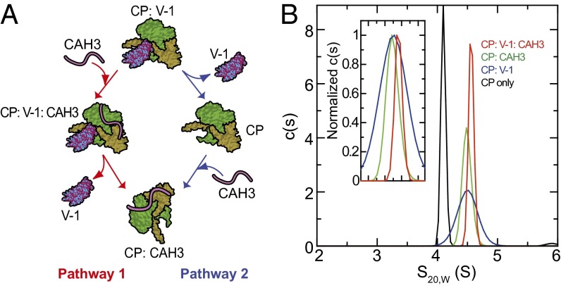
Although capping protein (CP) terminates actin filament elongation, it promotes Arp2/3-dependent actin network assembly and accelerates actin-based motility both in vitro and in vivo. In vitro, capping protein Arp2/3 myosin I linker (CARMIL) antagonizes CP by reducing its affinity for the barbed end and by uncapping CP-capped filaments, whereas the protein V-1/myotrophin sequesters CP in an inactive complex. Previous work showed that CARMIL can readily retrieve CP from the CP:V-1 complex, thereby converting inactive CP into a version with moderate affinity for the barbed end. Here we further clarify the mechanism of this exchange reaction, and we demonstrate that the CP:CARMIL complex created by complex exchange slows the rate of barbed-end elongation by rapidly associating with, and dissociating from, the barbed end. Importantly, the cellular concentrations of V-1 and CP determined here argue that most CP is sequestered by V-1 at steady state in vivo. Finally, we show that CARMIL is recruited to the plasma membrane and only at cell edges undergoing active protrusion. Assuming that CARMIL is active only at this location, our data argue that a large pool of freely diffusing, inactive CP (CP:V-1) feeds, via CARMIL-driven complex exchange, the formation of weak-capping complexes (CP:CARMIL) at the plasma membrane of protruding edges. In vivo, therefore, CARMIL should promote Arp2/3-dependent actin network assembly at the leading edge by promoting barbed-end capping there.

Keywords: VASP; cell migration.


Conflict of interest statement

The authors declare no conflict of interest.

Figures

Fig. 1.
Fig. 1.
CAH3-driven complex exchange can occur via two pathways, one of which involves the formation of a CP:V-1:CAH3 ternary complex. (A) Schematic of the two possible pathways by which CAH3 could drive the conversion of the CP:V-1 complex to the CP:CAH3 complex. (B) Data from analytical ultracentrifugation demonstrating the existence of the CP:V-1:CAH3 ternary complex. The c(s) distributions obtained for all four samples showed single dominant peaks in all cases representing over 95% of the loading concentrations, indicating that even in samples containing protein mixtures only single molecular species were detected. The c(s) distribution peak for the CP-only sample had a weight average s20,w value of 4.4 S and a calculated molar mass of 63.5 kDa, which is in good agreement with the theoretical molecular weight of CP. In the three samples containing protein mixtures, peaks representing free V-1 and CAH3 fractions were either not detectable or constituted less than 0.5% of the loading concentration. It is not possible to obtain reliable mass estimates for those complexes because the broadening of their sedimentation boundaries represent not only diffusion, but also complex exchange during sedimentation. The positions of the various complexes in the c(s) versus S-value plot are indicted by color coding (see key). (Inset) Normalized data.
Fig. 2.
Fig. 2.
The rate of spontaneous dissociation of V-1 from CP is accelerated dramatically by CAH3 via a mixture of pathways 1 and 2, with pathway 1 dominating at high concentrations of CAH3. (A) The rate of spontaneous dissociation of IAEDANS-(C2)-V-1 from a preformed complex of CP:IAEDANS-(C2)-V-1 (∼600 nM) in the presence of 60 µM unlabeled V-1, measured by fluorescence anisotropy. The estimated rate of spontaneous dissociation of IAEDANS-(C2)-V-1 from CP was k5 = 0.049 ± 0.015/s (Fig. S1B). (B) The rates of dissociation of IAEDANS-(C2)-V-1 from a preformed complex of CP:IAEDANS-(C2)-V-1 in the presence of 60 µM unlabeled V-1 and 0 (black), 25 nM (red) or 125 nM (blue) CAH3, measured by fluorescence anisotropy (data fit with single exponentials). (C) As in B, except that dissociation rates in assays containing more than 125 nM CAH3 were measured using a stopped-flow apparatus. The estimated equilibrium dissociation constant of CAH3 binding to CP:V-1 is Kd6 = 618 ± 164 nM, and the estimated rate of dissociation of V-1 from the CP:CAH3 complex is k7 = 3.57 ± 0.28/s (Fig. S1B). (D) The relative contribution of pathway 1 (ternary complex formation) to overall complex exchange (pathway 1 plus pathway 2) as a function of CAH3 concentration.
Fig. 3.
Fig. 3.
The CARMIL-1– and V-1–dependent regulatory cycle for CP and the affinity of V-1 for CP in the CP:CAH3 complex. (A) Proposed CARMIL-1– and V-1–dependent regulatory cycle for CP, based on data in this and previous studies (see Results). Step 1: V-1 inactivates a significant fraction of cellular CP by sequestering it in the CP:V-1 complex (barbed-end affinity = 0). Step 2: CARMIL drives via pathways 1 and 2 the conversion of the CP:V-1 complex into the CP:CARMIL complex, thereby partially activating CP (barbed-end affinity = ∼50 nM). Step 3: The CP:CARMIL complex caps the barbed end weakly. In contrast to free CP (barbed-end affinity = ∼0.1 nM), the CP:CARMIL complex allows barbed-end elongation, albeit at rates slower than for a free barbed end. Step 4: A factor and/or mechanism causes a large reduction in CARMIL’s affinity for CP, thereby driving the dissociation of CP from CARMIL. The free CP thus generated binds V-1 to repopulate the pool of sequestered CP. (B) The steady-state binding of a constant amount (200 nM) of IAEDANS-(MC7)-V-1 to increasing amounts (0–9 µM) of CP:CAH3 complex, estimated from the increase in the fluorescence of IAEDANS-(M7C)-V-1 that occurs upon binding, measured following 10 min of incubation at 22 °C. Of note, we could not distinguish the extent to which signal increase was from ternary complex formation or from IAEDANS-(M7C)-V-1 binding to CP following its dissociation from CAH3. That said, because V-1 dissociates very quickly from the ternary complex (3.6/s), and because CAH3 has a much higher affinity for both free CP and CP in the CP:V-1 complex than V-1 has for both free CP and CP in the CP:CAH3 complex, we assumed that the bulk of the increase in IAEDANS-(M7C)-V-1 fluorescence was due to its association in forming the ternary complex. The estimated equilibrium dissociation constant for the binding of IAEDANS-(M7C)-V-1 to the CP:CAH3 complex (Kd7) was 1.3 ± 0.3 µM (Fig. S1B).
Fig. 4.
Fig. 4.
CARMIL-driven complex exchange supports rates of barbed-end elongation in bulk solution assays that are in between those of free barbed ends and CP-capped ends. (A) Bulk actin polymerization assays performed using a solution containing ∼100 nM CP:V-1 complex, generated by incubating 100 nM CP with 6 µM V-1 for 10 min at 22 °C. Polymerization was then initiated by the addition of actin seeds (∼0.8 nM barbed ends) and 2 µM G-actin (10% pyrene-labeled). The black line shows the rate of polymerization in this control reaction. Forty seconds after initiating polymerization (arrow), we added to three parallel reactions a solution that represented 10% of the total volume of the reaction and that contained 0, 200 nM, or 2 µM CAH3, yielding final concentrations of 0 (green line), 20 nM (red line), or 200 nM (blue line) CAH3 (note: the gap following the arrow corresponds to the ∼20 s required to add CAH3 to the cuvette and return it to the fluorimeter). Note that the slight decrease in the rate for the green trace relative to the base rate (black trace) is due to the 10% dilution of the assay required for adding the solution with or without CAH3. (B) The initial rate of actin polymerization in solution assays containing 100 nM CP and 6 µM V-1 (i.e., ∼100 nM CP:V-1 complex) as a function of the amount of CAH3 present in the assay. Initial rates were estimated from a linear fitting of the increase in pyrene fluorescence during the first 40 s of the reaction. The values are expressed as a fraction of the control rate (0 nM CAH3).
Fig. 5.
Fig. 5.
Direct observation of the regulation of barbed-end elongation by the CP:CAH3 complex using single-filament imaging. (A) Representative time plots of the rate of growth in micrometers per second of individual actin filament barbed ends before the addition of the CP:CAH3 complex (to the left of the arrow marked “step 4” and preceding the plateau in elongation corresponding to the washing step, i.e., step 3), and after the addition of 0 nM (black), 25 nM (red), 100 nM (blue), or 200 nM (green) CP:CAH3 complex (to the right of the arrow marked “step 4”). (B) Kymographs showing the barbed-end elongation of single, representative actin filaments in the presence of 0 nM (Left) or 200 nM (Right) CP:CAH3 complex. (Scale bar, 5 µm.) (C) The fractional decrease in the rate of barbed-end elongation (relative to the rate in the absence of the CP:CAH3 complex) as a function of CP:CAH3 complex concentration. The data from TIRF assays are shown with black circles (Kd3 = 37 ± 5 nM), and the data from pyrene-based bulk solution assays are shown with red circles (Kd3 = 62 ± 11 nM). For TIRF assays, average barbed-end elongation rates were estimated from the linear fitting of 20 actin filaments for each complex concentration. For pyrene assays, each data point is the average of three independent experiments for each complex concentration (error bars represent the SD). We note that the inhibition of polymerization must also be due, to some small extent, to barbed-end capping by the very small amount of free CP that must be present in the assays. Such events should, however, be very rapidly reversed in our TIRF assays by CAH3-driven uncapping, given the very high concentration of CAH3 present in the assays (within less than 10 s; see figure 3 in ref. 14).
Fig. 6.
Fig. 6.
Quantitative analysis of the inhibition in growth rate of individual actin filament barbed ends by the CP:CAH3 complex. The black histograms show for each indicated CP:CAH3 complex concentration (upper right corner in boldface type) the change in length in intervals of 100 nm of individual actin filament barbed ends every 5 s, plotted as the fraction of total 5-s intervals scored (corresponding to the n value in upper right corner). The data at each complex concentration represent the behavior of at least 20 actin filaments. The n values rise as the complex concentration rises because the progressive slowing in growth rate associated with increasing complex concentration extends the time interval for accurately scoring length changes. Global fitting to two Gaussian distributions was performed (note that the peaks of the two distributions were not predefined). The Gaussian in red, which has a median rate of 0.23 µm/5 s, corresponds to the barbed end elongation phase, whereas the Gaussian in blue, which has a median rate of 0.00 µm/5 s, corresponds to the pause phase. The vertical dashed lines in red and blue mark the peaks of these two Gaussians.
Fig. 7.
Fig. 7.
mGFP-mCARMIL-1 localizes very close to, if not on, the plasma membrane. (A1) An overlaid image of an NRK cell cotransfected with mGFP-mCARMIL-1 and mCherry as a volume marker. The results of two separate intensity line scans (marked a and b in A1) in the green (mGFP-mCARMIL-1) and red (mCherry) channels are shown using green and red traces, respectively, in A2 (line scan a) and A3 (line scan b) (vertical black lines mark the cell boundary). (A2) Line scan a was at an edge undergoing active protrusion, and (A3) line scan b was at a stationary edge (based on the video sequences). (B1B7) Overlaid images of representative NRKs cotransfected with mCherry-mCARMIL-1 and GFP-actin (B1), GFP-Cortactin (B2), GFP-Cofilin (B3), GFP-Arp3 (B4), GFP-CPβ (B5), GFP-VASP (B6), or GFP-mCARMIL-1 (B7). A magnification of the boxed region in B6 is shown as separate red, green, and overlaid images at the bottom of that panel. (Scale bars, 5 µm.)
Fig. 8.
Fig. 8.
Distribution of endogenous mCARMIL-1 relative to other lamellipodia-resident proteins and the cell boundary. Representative immunofluorescence images of different primary MEFs stained for endogenous mCARMIL-1 (A), VASP (B), CP (D), or cortactin (E) or for F-actin (C) using phalloidin. (F) The distributions of fluorescence intensities for these four proteins and F-actin (color coded as indicated), obtained from intensity line scans beginning outside the cell and crossing into the cell (the cell boundary is indicated by the vertical black line). Each distribution represents the mean of 12–18 intensity line scans obtained from at least three cells. Colored arrowheads mark the peak of each intensity plot. (Scale bars, 5 µm.)
Fig. 9.
Fig. 9.
mGFP-mCARMIL-1 is recruited to the plasma membrane only at cell edges undergoing active protrusion and actin polymerization. (A1) A representative image of a PtK1 cell expressing GFP-actin (Movie S2). The arrow marks the position used to obtain the kymograph in A2. (A2) A kymograph for GFP-actin. The green boxes marked with a “P” in this and subsequent panels denote periods of edge protrusion (seen as positive slopes in the kymograph), and the red boxes marked with an “R” denote periods of edge retraction (seen as negative slopes in the kymograph). (B1) A representative image of a PtK1 cell expressing mGFP-mCARMIL-1 (Movie S3). Arrows 2–4 mark the positions used to obtain the kymographs in B2B4, respectively. (C1C3) A representative kymograph from a PtK1 cell coexpressing GFP-actin (C1) and mCherry-mCARMIL-1 (C2) (Movie S4). The overlaid image is shown in C3. (B5B8) Representative kymographs from PtK1 cells expressing GFP-VASP (B5) (Movie S5), GFP-CPβ (B6 and B7) (Movies S6 and S7), or GFP-cortactin (B8) (Movie S8). The time and distance scales for the kymographs in A2, B2B8, and C1C3 are indicated to the right of B8. (Scale bars: A1, 2 µm; B1, 5 µm.)

Similar articles

Cited by

References

    1. Pollard TD, Borisy GG. Cellular motility driven by assembly and disassembly of actin filaments. Cell. 2003;112(4):453–465. - PubMed
    1. Chhabra ES, Higgs HN. The many faces of actin: Matching assembly factors with cellular structures. Nat Cell Biol. 2007;9(10):1110–1121. - PubMed
    1. Campellone KG, Welch MD. A nucleator arms race: Cellular control of actin assembly. Nat Rev Mol Cell Biol. 2010;11(4):237–251. - PMC - PubMed
    1. Cooper JA, Sept D. New insights into mechanism and regulation of actin capping protein. Int Rev Cell Mol Biol. 2008;267:183–206. - PMC - PubMed
    1. Loisel TP, Boujemaa R, Pantaloni D, Carlier MF. Reconstitution of actin-based motility of Listeria and Shigella using pure proteins. Nature. 1999;401(6753):613–616. - PubMed