Review
. 2014 Dec;14(6):368-79.
doi: 10.1136/practneurol-2013-000758. Epub 2014 Apr 28.

The clinical approach to small fibre neuropathy and painful channelopathy

Affiliations
Review

The clinical approach to small fibre neuropathy and painful channelopathy

Andreas C Themistocleous et al. Pract Neurol. 2014 Dec.

Abstract

Small fibre neuropathy (SFN) is characterised by structural injury selectively affecting small diameter sensory and/or autonomic axons. The clinical presentation is dominated by pain. SFN complicates a number of common diseases such as diabetes mellitus and is likely to be increasingly encountered. The diagnosis of SFN is demanding as clinical features can be vague and nerve conduction studies normal. New diagnostic techniques, in particular measurement of intraepidermal nerve fibre density, have significantly improved the diagnostic efficiency of SFN. Management is focused on the treatment of the underlying cause and analgesia, as there is no neuroprotective therapy. A recent and significant advance is the finding that a proportion of cases labelled as idiopathic SFN are in fact associated with gain of function mutations of the voltage-gated sodium channels Nav1.7 and Nav1.8 (encoded by the genes SCN9A and SCN10A, respectively). There is a further group of heritable painful conditions in which gain of function mutations in ion channels alter excitability of sensory neurones but do not cause frank axon degeneration; these include mutations in Nav1.7 (causing erythromelalgia and paroxysmal extreme pain disorder) and TRPA1 (resulting in familial episodic pain disorder). These conditions are exceptionally rare but have provided great insight into the nociceptive system as well as yielding potential analgesic drug targets. In patients with no pre-existing risk factor, the investigation of an underlying cause of SFN should be systematic and appropriate for the patient population. In this review, we focus on how to incorporate recent developments in the diagnosis and pathophysiology of SFN into clinical practice.

Keywords: NEUROPATHY; PAIN.


Figures

Figure 1
Figure 1
Confocal images of skin biopsies taken from the legs of a control subject (A) and a patient with small fibre neuropathy secondary to HIV (B) showing PGP 9.5-immunoreactive fibres (red) and the basement membrane (labelled with type IV collagen fibres, green). Nerve fibres positive for PGP 9.5 (white arrows) are counted as they cross the dermal–epidermal junction. The intra-epidermal nerve fibres are absent in the patient with HIV (B) consistent with the diagnosis of small fibre neuropathy. Scale bar: 50 µm.
Figure 2
Figure 2
(A) Confocal image of a skin biopsy taken from the finger of a healthy subject illustrating different subtypes of sensory fibre: PGP 9.5 is used as an axonal marker (red) and myelin basic protein as a marker for myelin (green). There are numerous free nerve endings entering the epidermis (white arrows) and a single myelinated fibre is innervating a Meissner's corpuscle (yellow arrow). (B) Electron micrograph of the sural nerve. The blue arrow shows a large diameter myelinated fibre, the purple arrow marks a small diameter myelinated fibre and the red arrow marks a Remak bundle in which one non-myelinating Schwann cell associates with multiple unmyelinated C-fibres which are ensheathed in Schwann cell pockets.
Figure 3
Figure 3
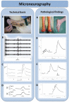
Microneurography in humans: microneurography is an electrophysiological technique to record action potentials from individual peripheral nerve axons in humans. Technical basis (left). (A) Microneurography setting. A tungsten microelectrode (1) is inserted intraneurally in a peripheral nerve; in this case the superficial peroneal nerve at the dorsum of the foot. A subcutaneous reference electrode (2) is inserted in the skin outside the nerve trunk. The innervation territory of the nerve is electrically stimulated with a pair of needle electrodes (3). In this example, conduction distance between stimulating electrodes and an active microneurography electrode is 100 mm. (B) Electrophysiological recording of responses. Electrical stimulation of the receptive field evokes multiple action potentials in the sweep. Latencies of these action potentials allow segregation of units between a group of thinly myelinated, Aδ units (latencies ≈ 10 ms; conduction velocity ≈ 10 m/s) and a group of unmyelinated C-units (latencies ≈ 100 ms; conduction velocities ≈ 1 m/s). Superposition of the individual sweeps in a cascade mode demonstrates reproducibility of the responses. (C) Raster plot of latencies. The individual sweeps displayed in a cascade mode in B can also be displayed as a raster plot of latencies, with latency as the y-axes and elapsed time as the x-axes. In this example, three C-units at approximate latencies of 140, 155 and 180 ms at min 40 are displayed. Unmyelinated C axons change their conduction velocity (and hence their latency) depending on the stimulation frequency, a phenomenon called ‘activity-dependent slowing of conduction velocity’. For example, baseline stimulation at 0.25 Hz can be interrupted (open bar) or increased to 2 Hz (filled bar). This induces changes in latency with different profiles. (D) Profiles of activity-dependent slowing of conduction velocity. Different subpopulations of peripheral unmyelinated axons display different profiles of conduction slowing. For example, C-nociceptors have characteristic ‘shark fin’ profiles. Pathological findings (right). (A) Neuropathic pain. Peripheral nerve damage, such as an axonal polyneuropathy (left) or a posterior tibial nerve damage (right) frequently induce positive sensory phenomena. Burning or deep aching pain, due to activity arising from C-nociceptors, is a frequent complaint in neuropathic pain patients. (B) Spontaneous discharges in C-nociceptors. Damaged C-nociceptors frequently engage in ongoing, spontaneous activity. Spontaneous discharges are indicated by abrupt increases in latency (black arrows) between successive electrical stimuli delivered at 0.25 Hz, giving rise to a characteristic ‘saw-tooth’ profile of the raster plot. (C) Peripheral sensitisation in C-nociceptors. Example of mechanically evoked activity in a normally mechano-insensitive C-nociceptor recorded from a patient suffering from small-fibre neuropathy. Spontaneous discharges are evoked by pressing with a von Frey hair exerting a force of 128 mN (arrow). (D) Double spike in pathological C-nociceptor. Under normal conditions, single electrical shocks into the receptive field of C-nociceptors induce single action potentials. In neuropathy, with frequent dying-back and regeneration attempts, extensive, abnormal branchings may induce ‘unidirectional blocks’ in the terminal arborisation of the units, and induce multiple spikes (arrows signal beginning and end of the double spike), thus amplifying peripheral input. Double and triple spikes may contribute to hyperalgesia in patients with neuropathic pain.
Figure 4
Figure 4
An example of a quantitative sensory testing profile for a patient with small fibre neuropathy. Data are reported as z-score profiles for each sensory test as depicted here. Z-score is defined as the SD of the recorded result from the mean normal data result and calculated as z=(value patient−mean controls)/SDcontrols. Each data point is discrete; however, they are connected for graphical illustration as a z profile. Quantitative sensory tests included CDT, cold detection threshold; WDT, warm detection threshold; TSL, thermal sensory limen; CPT, cold pain threshold; HPT, heat pain threshold; PPT, pressure pain threshold; MPT, mechanical pain threshold; MPS, mechanical pain sensitivity; WUR, wind up ratio; MDT, medical detection threshold; and VDT, vibration detection threshold. Normal data are distributed within the shaded area (mean at 0±2 SD). Note selective and significant impairment of small fibre function including hyposensitivity to cooling, warming, elevated heat pain threshold and impaired thermal sensory limen.
Figure 5
Figure 5
Algorithm for diagnosis of small fibre neuropathy.
Figure 6
Figure 6
Photograph of the legs of a patient with inherited erythromelalgia, showing erythema to the level of the mid-calf. With increasing ambient temperature, this erythema becomes more extensive. (Reproduced with permission of the International Association for the Study of Pain (IASP) from Segerdahl et al).

References

    1. Hoeijmakers JG, Faber CG, Lauria G, et al. Small-fibre neuropathies-advances in diagnosis, pathophysiology and management. Nat Rev Neurol 2012;8:369–79. - PubMed
    1. Hovaguimian A, Gibbons CH. Diagnosis and treatment of pain in small-fiber neuropathy. Curr Pain Headache Rep 2011;15:193–200. - PMC - PubMed
    1. Waxman SG. Painful Na-channelopathies: an expanding universe. Trends Mol Med 2013;19:406–9. - PubMed
    1. Kremeyer B, Lopera F, Cox JJ, et al. A gain-of-function mutation in TRPA1 causes familial episodic pain syndrome. Neuron 2010;66:671–80. - PMC - PubMed
    1. Devigili G, Tugnoli V, Penza P, et al. The diagnostic criteria for small fibre neuropathy: from symptoms to neuropathology. Brain 2008;131:1912–25. - PMC - PubMed

Publication types

MeSH terms