. 2014 Jun 1;192(11):5214-25.
doi: 10.4049/jimmunol.1302806. Epub 2014 Apr 28.

Longitudinal requirement for CD4+ T cell help for adenovirus vector-elicited CD8+ T cell responses

Affiliations

Longitudinal requirement for CD4+ T cell help for adenovirus vector-elicited CD8+ T cell responses

Nicholas M Provine et al. J Immunol. .

Abstract

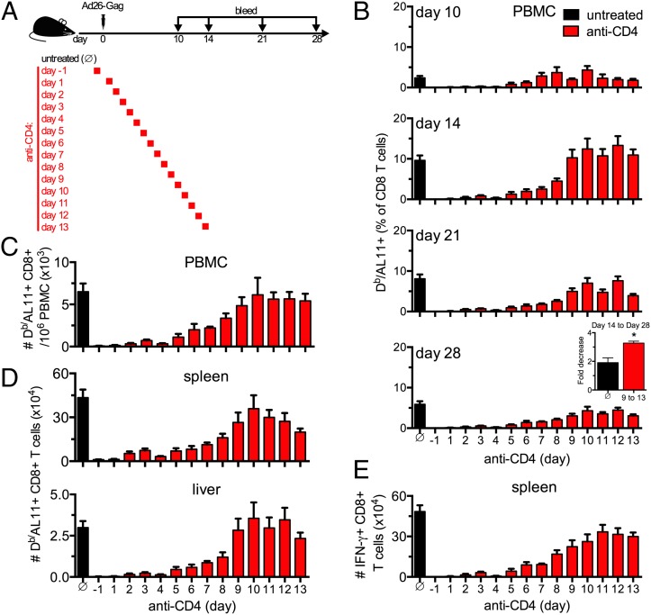
Despite the widespread use of replication-incompetent recombinant adenovirus (Ad) vectors as candidate vaccine platforms, the mechanism by which these vectors elicit CD8(+) T cell responses remains poorly understood. Our data demonstrate that induction and maintenance of CD8(+) T cell responses by Ad vector immunization is longitudinally dependent on CD4(+) T cell help for a prolonged period. Depletion of CD4(+) T cells in wild type mice within the first 8 d following Ad immunization resulted in dramatically reduced induction of Ag-specific CD8(+) T cells, decreased T-bet and eomesodermin expression, impaired KLRG1(+) effector differentiation, and atypical expression of the memory markers CD127, CD27, and CD62L. Moreover, these CD8(+) T cells failed to protect against a lethal recombinant Listeria monocytogenes challenge. Depletion of CD4(+) T cells between weeks 1 and 4 following immunization resulted in increased contraction of memory CD8(+) T cells. These data demonstrate a prolonged temporal requirement for CD4(+) T cell help for vaccine-elicited CD8(+) T cell responses in mice. These findings have important implications in the design of vaccines aimed at eliciting CD8(+) T cell responses and may provide insight into the impaired immunogenicity of vaccines in the context of AIDS and other CD4(+) T cell immune deficiencies.


Figures

FIGURE 1.
FIGURE 1.
CD4+ T cell help is required for the development of CD8+ T cell responses following i.m. Ad vaccination. (A and B) C57BL/6, CD4 KO, or MHC class II KO mice were immunized i.m. with 109 vp of Ad26-Gag or were unimmunized (naive C57BL/6). Animals were bled longitudinally, and SIV Gag-specific CD8+ T cells were quantified by tetramer staining. Representative plots (A) and group averages (B) for Db/AL11+ cells as a percent of CD8+ T cells are shown. Data are from n = 8 per group (pooled from two independent experiments). (C and D) C57BL/6, CD4 KO, or MHC class II KO mice were coimmunized i.m. with 109 vp each of Ad26-Gag and Ad26-Env. Splenocytes were harvested on day 28 after immunization and stimulated with the indicated peptide pool. Representative plots of IFN-γ+ CD8+ T cells (C) and group averages (D) are shown. Data are from n = 8 per group (pooled from two independent experiments). (E) C57BL/6, CD4 KO, or MHC class II KO mice were coimmunized i.m. with 109 vp each of Ad5HVR48-Gag and Ad5HVR48-Env. Splenocytes were harvested on day 28 after immunization and stimulated with the indicated peptide pool. Percent of IFN-γ+ cells as a fraction of CD8+ T cells are shown. Data are from n = 8 per group (pooled from two independent experiments). Mean ± SEM are shown. *p < 0.01.
FIGURE 2.
FIGURE 2.
Temporal requirement for CD4+ T cell help for the induction and expansion of CD8+ T cell responses following Ad vaccination. (A) C57BL/6 mice were depleted of CD4+ T cells by i.p. administration of mAb GK1.5 (anti-CD4) beginning on days −1, 1, 2, 3, 4, 5, 6, 7, 8, 9, 10, 11, 12, or 13 and immunized i.m. with 109 vp of Ad26-Gag on day 0. Untreated controls are designated by a circle with a diagonal line though it. (B) Mice from each treatment group were bled at the indicated time points, and AL11-specific CD8+ T cells were assessed by Db/AL11 staining. Inset, the fold-decrease in the number of AL11-specific CD8+ T cells in blood from day 14 to 28 after immunization is shown. Groups of mice administered with anti-CD4 beginning on day 9–13 were pooled for analysis. (C and D) On day 28 after immunization, the total number of AL11 peptide-specific CD8+ T cells per 106 PBMCs (C) or per tissue (D) were quantified by Db/AL11 staining. (E) On day 28 after immunization, the total number of AL11 peptide-specific CD8+ T cells was assessed by ex vivo restimulation with AL11 peptide followed by intracellular cytokine staining for IFN-γ. Data are from n = 7–22 per group (pooled from two to six independent experiments). Mean ± SEM are shown. *p < 0.001.
FIGURE 3.
FIGURE 3.
Effector differentiation of CD8+ T cells is dependent on CD4+ T cell help following Ad vaccination. C57BL/6 mice were depleted of CD4+ T cells by i.p. administration of mAb GK1.5 (anti-CD4) beginning on days 4, 5, 6, 7, 8, 9, 10, 11, 12, or 13 or were left untreated, and were immunized i.m. with 109 vp of Ad26-Gag on day 0. On day 28 after immunization, animals were sacrificed and tissues were harvested. KLRG1 and CD127 expression on Db/AL11+ CD8+ T cells was assessed. (A and B) Representative flow plots (A) and group averages (B) of KLRG1 and CD127 expression on Db/AL11+ CD8+ T cells in the spleen. (C and D) Group summary of KLRG1 and CD127 expression of Db/AL11+ CD8+ T cells in blood (C) and liver (D). (E) Absolute number of KLRG1+CD127 or KLRG1CD127+ Db/AL11+ CD8+ T cells in the spleen. Anti-CD4 treatment on days −1, 1, 2, and 3 after immunization had insufficient numbers of Db/AL11+ cells for analysis. Data are from n = 8–16 per group (pooled from two to four independent experiments), except for PBMC anti-CD4 day 9, 10, and 12 groups, which are n = 3–4 per group (one experiment). Mean ± SEM are shown. *p < 0.05.
FIGURE 4.
FIGURE 4.
CD8+ T cells in mice depleted of CD4+ T cells exhibit an abnormal phenotype after i.m. Ad vaccination. C57BL/6 mice were depleted of CD4+ T cells by i.p. administration of mAb GK1.5 (anti-CD4) on days 5, 8, or 11 or left untreated and immunized i.m. with 109 vp of Ad26-Gag. On day 28 after immunization, animals were sacrificed and splenocytes were isolated. (A and B) Expression level of the indicated protein as a percentage (A) or mean fluorescence intensity (B) on gated Db/AL11+ cells. Dashed lines indicate CD44lo CD8+ (naive) T cells, and solid-shaded histograms indicate Db/AL11+ CD8+ T cells. Data are from n = 8–16 per group (pooled from two to four independent experiments). (C) Gene expression of sorted Db/AL11+ CD8+ T cells from untreated animals or animals treated with GK1.5 (anti-CD4) on days 5 or 8 after immunization was determined by quantitative real-time PCR. Gene expression was normalized to Gapdh. Data are from n = 4–6 per group (pooled from two independent experiments). (D) Annexin V expression and uptake of vital dye by Db/AL11+ cells was assessed. Data are from n = 8 per group (pooled from two independent experiments). Mean ± SEM are shown. #p = 0.067, *p < 0.05.
FIGURE 5.
FIGURE 5.
CD8+ T cells primed with reduced CD4+ T cell help fail to protect against lethal recombinant L. monocytogenes challenge. (A) C57BL/6 mice were depleted of CD4+ T cells by i.p. administration of mAb GK1.5 (anti-CD4) on day 8 or left untreated, and immunized with 109 vp of Ad26-GP. On day 30 after immunization, mice were challenged with 1.75 × 10⁵ to 2.5 × 10⁵ CFU Lm-GP33. (B) Frequency of Db/GP33+ CD8+ T cells in the blood on day 30 after immunization. (C) KLRG1 and CD127 expression on Db/GP33+ CD8+ T cells in the blood on day 30 after immunization. (D) Survival of mice after Lm-GP33 challenge. Data are from n = 16–32 per group (A–C) or n = 7–19 per group (D) pooled from two to four independent experiments. Mean ± SEM are shown. #p = 0.08, *p < 0.05.
FIGURE 6.
FIGURE 6.
Normal contraction of the CD8+ T cell response requires prolonged CD4+ T cell help. (A) C57BL/6 mice were depleted of CD4+ T cells by i.p. administration of mAb GK1.5 (anti-CD4) on days −1, 7, 14, 21, or 28 or left untreated, and immunized i.m. with 109 vp of Ad26-Gag. (B) Animals were bled longitudinally, and Gag-specific CD8+ T cells were quantified by Db/AL11 tetramer staining. (C) Frequency of KLRG1 and CD127 expression on Db/AL11+ CD8+ T cells in blood. Data are from n = 8–12 per group (pooled from two to three independent experiments). Mean ± SEM are shown. *p < 0.05.
FIGURE 7.
FIGURE 7.
CD8+ T cells primed with at least 8 d of CD4+ T cell help proliferate upon secondary Ag exposure. (A) CD45.1+ mice were depleted of CD4+ T cells by i.p. administration of mAb GK1.5 (anti-CD4) on days 8, 11, or 14 or left untreated, and immunized i.m. with 109 vp of Ad26-Gag (day 0). On day 28 postimmunization, CD8+ T splenocytes were pooled, enriched by negative selection, and labeled with CFSE; 1.5 × 10⁴ Db/AL11+ CD8+ T cells were transferred by tail vein injection into naive CD45.2+ recipients. One day after transfer, animals were immunized i.m. with 109 vp of Ad5HVR48-Gag. (B) Representative plots of Db/AL11+ CD45.1+ donor cells as a fraction of CD8+ T cells in the spleen on days 8 or 14. (C) Representative histograms of CFSE dilution in the donor Db/AL11+ CD45.1+ CD8+ T cell population on day 8 after boost (open histogram) or the donor CD45.1+ CD8+ T cell population with no boosting immunization (solid histogram). (D) Absolute number of Db/AL11+ CD45.1+ CD8+ T cells in the spleen on days 8 or 14 after immunization. Frequency of donor Db/AL11+ CD45.1+ CD8+ T cells at the time of immunization (day 0) is based on 10% engraftment. Data are from n = 6–13 recipient mice per group on day 8 (pooled from three independent experiments) and n = 5 recipient mice per group on day 14 (from one experiment). Mean ± SEM are shown.
FIGURE 8.
FIGURE 8.
CD40 signaling is required for programming CD8+ T cells with anamnestic potential. (AC) C57BL/6, CD40L KO, and CD40 KO animals were immunized i.m. with 109 vp of Ad26-Gag (n = 8 per group pooled from two independent experiments). (A) The number of Db/AL11+ CD8+ T cells in the spleen of immunized animals on day 28 after immunization. (B) Splenocytes were harvested on day 28 after immunization and the number of IFN-γ+ CD8+ T cells was quantified following stimulation with an overlapping SIV Gag peptide pool. (C) KLRG1 and CD127 expression were assessed on splenic Db/AL11+ CD8+ T cells on day 28 after immunization. (D and E) C57BL/6, CD40L KO, and CD40 KO animals were immunized i.m. with 109 vp of Ad26-Gag and on day 56 after priming were boosted with 109 vp of Ad5HVR48-Gag (n = 4 per group from one experiment). (D) The frequency of Db/AL11+ CD8+ T cells per 106 PBMCs after the boost was assessed. (E) The fold-change in frequency of Db/AL11+ CD8+ T cells per 106 PBMCs from 1 d before boost to day 7 after boost was assessed. (F) C57BL/6 or CD40L KO animals were immunized i.m. with 109 vp of Ad26-Gag. On day 50 after immunization, 5 × 10⁴ Db/AL11+ CD8+ T cells were enriched by negative selection and transferred i.v. to congenically marked (CD45.1+) naive recipients (n = 8–9 per group from two independent experiments). One day after transfer, recipient mice were immunized i.m. with 109 vp of Ad5HVR48-Gag. On day 12 after secondary immunization, the number of donor Db/AL11+ CD45.2+ CD8+ T cells in PBMCs, spleen, and liver was assessed. Mean ± SEM are shown. #p = 0.06, *p < 0.05.
FIGURE 9.
FIGURE 9.
Schematic representation of longitudinal requirement for CD4+ T cell help. Summary of longitudinal requirement for CD4+ T cell help for the induction of CD8+ T cell responses, effector differentiation, and normal contraction following Ad vector vaccination. The requirement for CD4+ T cell help was prolonged but waned over time.

Similar articles

Cited by

References

    1. Sullivan N. J., Hensley L., Asiedu C., Geisbert T. W., Stanley D., Johnson J., Honko A., Olinger G., Bailey M., Geisbert J. B., et al. 2011. CD8+ cellular immunity mediates rAd5 vaccine protection against Ebola virus infection of nonhuman primates. Nat. Med. 17: 1128–1131 - PubMed
    1. Alexander J., Ward S., Mendy J., Manayani D. J., Farness P., Avanzini J. B., Guenther B., Garduno F., Jow L., Snarsky V., et al. 2012. Pre-clinical evaluation of a replication-competent recombinant adenovirus serotype 4 vaccine expressing influenza H5 hemagglutinin. PLoS ONE 7: e31177. - PMC - PubMed
    1. Barnes E., Folgori A., Capone S., Swadling L., Aston S., Kurioka A., Meyer J., Huddart R., Smith K., Townsend R., et al. 2012. Novel adenovirus-based vaccines induce broad and sustained T cell responses to HCV in man. Sci. Transl. Med. 4: 115ra111 - PMC - PubMed
    1. Colloca S., Barnes E., Folgori A., Ammendola V., Capone S., Cirillo A., Siani L., Naddeo M., Grazioli F., Esposito M. L., et al. 2012. Vaccine vectors derived from a large collection of simian adenoviruses induce potent cellular immunity across multiple species. Sci. Transl. Med. 4: 115ra112 - PMC - PubMed
    1. Xiang Z. Q., Yang Y., Wilson J. M., Ertl H. C. 1996. A replication-defective human adenovirus recombinant serves as a highly efficacious vaccine carrier. Virology 219: 220–227 - PubMed

Publication types

MeSH terms