. 2014 Jun 1;192(11):5296-304.
doi: 10.4049/jimmunol.1400653. Epub 2014 Apr 28.

Endothelial PINK1 mediates the protective effects of NLRP3 deficiency during lethal oxidant injury

Affiliations

Endothelial PINK1 mediates the protective effects of NLRP3 deficiency during lethal oxidant injury

Yi Zhang et al. J Immunol. .

Abstract

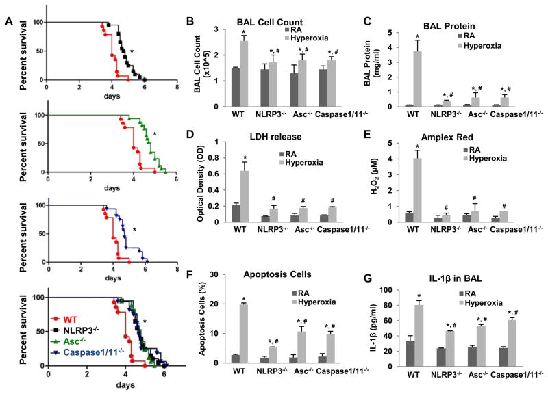
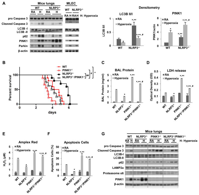
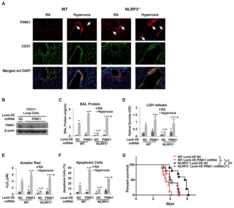
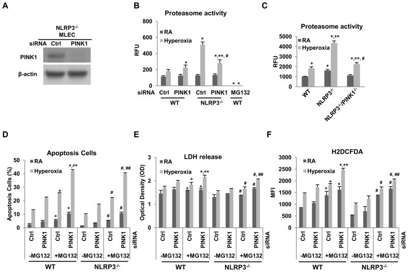
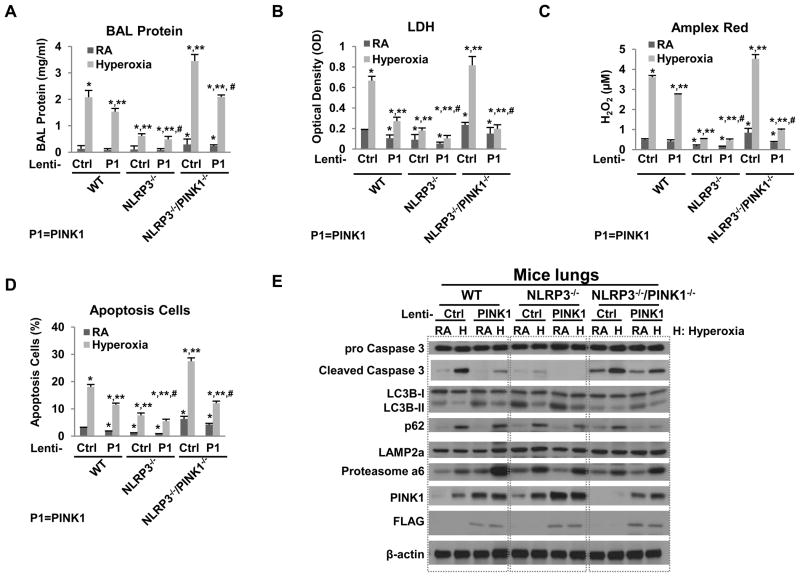
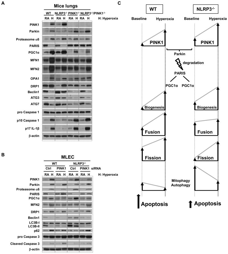
High levels of inspired oxygen, hyperoxia, are frequently used in patients with acute respiratory failure. Hyperoxia can exacerbate acute respiratory failure, which has high mortality and no specific therapies. We identified novel roles for PTEN-induced putative kinase 1 (PINK1), a mitochondrial protein, and the cytosolic innate immune protein NLRP3 in the lung and endothelium. We generated double knockouts (PINK1(-/-)/NLRP3(-/-)), as well as cell-targeted PINK1 silencing and lung-targeted overexpression constructs, to specifically show that PINK1 mediates cytoprotection in wild-type and NLRP3(-/-) mice. The ability to resist hyperoxia is proportional to PINK1 expression. PINK1(-/-) mice were the most susceptible; wild-type mice, which induced PINK1 after hyperoxia, had intermediate susceptibility; and NLRP3(-/-) mice, which had high basal and hyperoxia-induced PINK1, were the least susceptible. Genetic deletion of PINK1 or PINK1 silencing in the lung endothelium increased susceptibility to hyperoxia via alterations in autophagy/mitophagy, proteasome activation, apoptosis, and oxidant generation.


Figures

Figure 1
Figure 1. NLRP3−/−, Asc−/− and Caspase 1/11−/− mice are more resistant to hyperoxia
WT, NLRP3−/−, Asc−/− and Caspase 1/11−/− mice were exposed to continuous hyperoxia (A) or 72h of hyperoxia (B–G). RA, room air control. Survival proportions were compared among each set or four groups (A). *p <0.05 vs WT mice (n=15~18 for each group). B) Lung inflammation was detected by BAL cell counts. C) Lung permeability was assessed by BAL protein content. D) LDH activity assay from BAL fluid. E) Oxidant generation was detected by Amplex red from BAL fluid. F) TUNEL staining was performed on lung sections and the number of TUNEL-positive cells were quantitated and expressed as a percentage of the total number of lung cells counted on each section. G) IL1β was detected by ELISA in BAL. The values are expressed as mean ± SD. *p <0.05 vs RA WT mice; #p<0.05 vs hyperoxia WT mice (n=5 for each group).
Figure 2
Figure 2. PINK1 is protective in WT and NLRP3−/− mice
A) Mice or MLEC were exposed to RA or 72h hyperoxia. Lysates from mouse lungs or MLEC were isolated and immunoblotted against antibodies as listed. β-actin was used as protein loading control. RA, room air control; H, hyperoxia exposure. The left panel shows a representative Western blot, and the right panel shows the quantification based on densitometry for LC3B II/I and PINK to β-actin (experiments were performed in triplicate). B) Survival was compared amongst WT, NLRP3−/−, PINK1−/− and NLRP3−/−/PINK1−/− mice. *p <0.05 vs WT mice; #p <0.05 vs PINK1−/− mice, ##p <0.05 vs NLRP3−/− mice (n=10 for each group). C~F) WT, NLRP3−/−, PINK1−/− and NLRP3−/−/PINK1−/− mice were exposed to 72h of hyperoxia. C) Lung permeability. D) LDH activity. E) Oxidant generation. F) TUNEL-positive cells percentage. The values are expressed as mean ± SD. *p<0.05 vs WT mice; **p< 0.05 vs NLRP3−/− mice; #p<0.05 vs PINK1−/− mice (n=5 in each group). G) Lysates from mouse lungs were isolated and immunoblotted against antibodies as listed. One representative western blot out of three experiments is shown.
Figure 3
Figure 3. Lung endothelial-targeted PINK1 knockdown increased susceptibility to hyperoxia in vivo
A) Co-localization of PINK1 and endothelium in lung sections using immunofluorescence. Mice were exposed to RA or 72h hyperoxia. Lung sections were immunostained for PINK1 (red) and CD31 (green). Nuclei were stained with DAPI (blue). Upper panels show single immunostained images; lower panels show merged images. Original view of all photomicrographs × 200. Arrows indicate the PINK1 positive cells. The results are representative of at least three independent experiments. WT and NLRP3−/− mice were treated with intranasal lentivirus (lenti-VE NC or lenti-VE PINK1 miRNA) and exposed to RA or 72h hyperoxia. B) Cell lysates of lung endothelium immunoselected by α-CD31 antibody from NLRP3−/− lungs were immunoblotted against PINK1 antibody. β-actin was used as protein loading control. One representative western blot out of three experiments is shown. C) Lung permeability. D) LDH activity. E) Oxidant generation. F) TUNEL-positive cells percentage. The values are expressed as mean ± SD.*p<0.05 vs RA lenti-VE NC WT mice; **p< 0.05 vs hyperoxia lenti-VE NC WT mice; #p<0.05 vs hyperoxia lenti-VE PINK1 miRNA WT mice (n=5 in each group). G) WT and NLRP3−/− mice were administered intranasal lentivirus for two weeks and exposed to continuous hyperoxia. Survival was compared amongst four groups. *p <0.05 vs WT lenti-VE NC mice; #p <0.05 vs NLRP3−/− lenti-VE NC mice; ##p<0.05 vs NLRP3−/− lenti-VE PINK1 miRNA mice (n=10 for each group).
Figure 4
Figure 4. NLRP3−/− had higher proteasome activity
A) Lysates from NLRP3−/− MLECs transfected with PINK1 siRNA or Ctrl siRNA and immunoblotted against PINK1 antibody. β-actin was used as protein loading control. One representative western blot out of three experiments is shown. Proteasome activity was measured in MLEC transfected with PINK1 siRNA (B) or in lungs of WT, NLRP3−/− and NLRP3−/−/PINK1−/− mice (C) exposed to 72h of hyperoxia. MLEC treated with proteasome inhibitors, MG-132 at 10 μM, was used as a negative control. The values are expressed as mean ± SD. *p<0.05 vs corresponding WT Ctrl siRNA; **p<0.05 vs corresponding WT PINK1 siRNA; #p<0.05 vs corresponding NLRP3−/− in (B); *p<0.05 vs WT RA; **p<0.05 vs WT hyperoxia; #p<0.05 vs NLRP3−/− hyperoxia (n=5 for each group) in (C). WT and NLRP3−/− MLEC were transfected with PINK1 siRNA and treated with proteasome inhibitor MG-132 at 10μM (D~F), and then exposed to RA or 72h hyperoxia. D) Graphical quantitation of flow cytometry analysis of apoptosis. E) LDH activity assay from MLEC supernatant. F) H2O2 generation by CM-H2DCFDA staining. The values are expressed as mean ± SD. *p <0.05 vs corresponding group in no-MG132 WT MLEC; **p <0.05 vs corresponding Ctrl siRNA in WT; #p<0.05 vs corresponding group in no-MG132 NLRP3−/− MLEC; ##p<0.05 vs corresponding Ctrl siRNA in NLRP3−/− (experiments were performed in triplicates).
Figure 5
Figure 5. Lung-targeted PINK1 overexpression decreased susceptibility to hyperoxia in vivo
WT, NLRP3−/− and NLRP3−/−/PINK1−/− mice were administered intranasal lentivirus (lenti-Ctrl or lenti-PINK1) and exposed to RA or 72h hyperoxia. A) Lung permeability. B) LDH activity. C) Oxidant generation. D) TUNEL-positive cells percentage. The values are expressed as mean ± SD.*p<0.05 vs RA lenti-Ctrl WT mice; **p< 0.05 vs hyperoxia lenti-Ctrl WT mice; #p<0.05 vs hyperoxia lenti- PINK1 WT mice (n=5 in each group). E) Lysates from mouse lungs were isolated and immunoblotted against antibodies as listed. One representative western blot out of three experiments is shown.
Figure 6
Figure 6. NLRP3 deficiency promotes mitochondrial maintenance and autophagy
A) Lysates from mouse lungs were immunoblotted against antibodies as listed. B) WT and NLRP3−/− MLEC were transfected with PINK1 siRNA. Lysates from MLEC were immunoblotted against antibodies as listed. β-actin was used as protein loading control. RA, room air control; H, hyperoxia exposure. One representative western blot out of three experiments is shown. C) Proposed schematic. Mitochondria proliferate from preexisting mitochondria and generate new mitochondria (biogenesis). PINK1 recruits Parkin to the mitochondria and eliminates damaged mitochondria through autophagy/mitophagy. Parkin ubiquitinates and degrades PARIS through ubiquitin-proteasome pathway (USP). In NLRP3 deficiency, PINK1-Parkin is induced, PARIS is degraded and mitochondrial biogenesis is activated via PGC-1α. NLPR3 deficiency or PINK1 induction leads to reduced apoptosis. The arrows indicate the direction of change for each of the cellular processes; the height of the arrows indicates relative degree of change.

References

    1. Fisher AB. Oxygen therapy. Side effects and toxicity. Am Rev Respir Dis. 1980;122:61–69. - PubMed
    1. Fukumoto J, Fukumoto I, Parthasarathy PT, Cox R, Huynh B, Ramanathan GK, Venugopal RB, Allen-Gipson DS, Lockey RF, Kolliputi N. NLRP3 deletion protects from hyperoxia-induced acute lung injury. Am J Physiol Cell Physiol. 2013;30:C182–189. - PMC - PubMed
    1. Matsuda S, Kitagishi Y, Kobayashi M. Function and characteristics of PINK1 in mitochondria. Oxid Med Cell Longev. 2013;2013:601587. - PMC - PubMed
    1. Sutterwala FS, Ogura Y, Szczepanik M, Lara-Tejero M, Lichtenberger GS, Grant EP, Bertin J, Coyle AJ, Galan JE, Askenase PW, Flavell RA. Critical role for NALP3/CIAS1/Cryopyrin in innate and adaptive immunity through its regulation of caspase-1. Immunity. 2006;24:317–327. - PubMed
    1. Kuida K, Lippke JA, Ku G, Harding MW, Livingston DJ, Su MS, Flavell RA. Altered cytokine export and apoptosis in mice deficient in interleukin-1 beta converting enzyme. Science. 1995;267:2000–2003. - PubMed

Publication types

MeSH terms