Comparative evaluation of immunohistochemistry, histopathology and conventional radiography in differentiating periapical lesions
- PMID: 24778515
- PMCID: PMC4001275
- DOI: 10.4103/0972-0707.128061
Comparative evaluation of immunohistochemistry, histopathology and conventional radiography in differentiating periapical lesions
Abstract
Background and aim: Periapical lesions often present differently on the radiograph resulting in a dilemma in the mind of the dentist to arrive at a final diagnosis. Although, histopathologic diagnosis has been used for confirmation of the true nature of periapical lesion, the concept of transformation of periapical granulomas containing epithelium without cystification into cyst remains controversial. The aim of this in vivo study was to evaluate the efficacy of conventional radiography and histopathology in differentiating periapical lesions in adjunct with immunohistochemical analysis.
Aim: Periapical lesions often present differently on the radiograph resulting in a dilemma in the mind of the dentist to arrive at a final diagnosis. Although, histopathologic diagnosis has been used for confirmation of the true nature of periapical lesion, the concept of transformation of periapical granulomas containing epithelium without cystification into cyst remains controversial. The aim of this in vivo study was to evaluate the efficacy of conventional radiography and histopathology in differentiating periapical lesions in adjunct with immunohistochemical analysis.
Materials and method: Thirty patients having large periapical radiolucency that do not heal successfully with routine endodontic therapy in relation to either maxillary or mandibular anterior teeth were selected for the study. Intraoral periapical radiographs were obtained and provisional diagnosis of the apical areas were made. Endodontic surgery was performed to enable histopathogical investigation. The histopathological interpretation was done to arrive at a final diagnosis and selected questionable granulomas were subjected for cytokeratin (CK-14) stain.
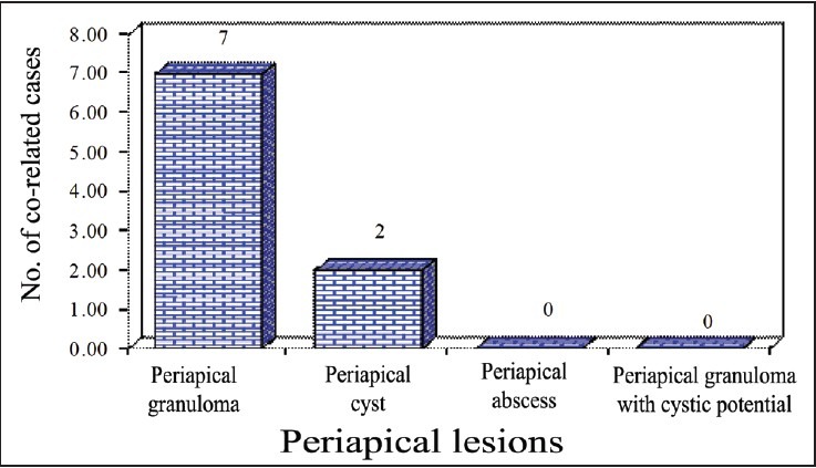
Results: The histopathological profile of lesions consisted of 66.66% periapical granulomas, 10% cysts, 6.67% abscess and 16.67% granulomas with cystic potential. The radiographic and histopathologic correlation was found in only 30% of these cases. Strong CK-14 expression was observed in all five cases of periapical granuloma with cystic potential.
Conclusion: The radiographic diagnosis of periapical lesions remains inconclusive. Although histopathologic examination of periapical lesions gives true nature, the precise nature of subsets of periapical granulomas may be achieved with adjunct use of immunohistochemical markers.
Keywords: Conventional radiography; Cytokeratin-14; Periapical lesions.
Conflict of interest statement
Figures


Similar articles
-
Ultrasonography with color Doppler and power Doppler in the diagnosis of periapical lesions.Indian J Radiol Imaging. 2011 Oct;21(4):279-83. doi: 10.4103/0971-3026.90688. Indian J Radiol Imaging. 2011. PMID: 22223940 Free PMC article.
-
Comparison of ultrasound, digital and conventional radiography in differentiating periapical lesions.Dentomaxillofac Radiol. 2006 Sep;35(5):326-33. doi: 10.1259/dmfr/60326577. Dentomaxillofac Radiol. 2006. PMID: 16940480
-
A Comparative Evaluation of Digital Radiography and Ultrasound Imaging to Detect Periapical Lesions in the Oral Cavity.Cureus. 2022 Oct 8;14(10):e30070. doi: 10.7759/cureus.30070. eCollection 2022 Oct. Cureus. 2022. PMID: 36381877 Free PMC article.
-
Nonsurgical root canal therapy of large cyst-like inflammatory periapical lesions and inflammatory apical cysts.J Endod. 2009 May;35(5):607-15. doi: 10.1016/j.joen.2009.02.012. J Endod. 2009. PMID: 19410070 Review.
-
A dentist's dilemma: nonsurgical endodontic therapy or periapical surgery for teeth with apparent pulpal pathosis and an associated periapical radiolucent lesion.Oral Surg Oral Med Oral Pathol. 1990 Sep;70(3):333-40. doi: 10.1016/0030-4220(90)90151-h. Oral Surg Oral Med Oral Pathol. 1990. PMID: 2216362 Review.
Cited by
-
Apical periodontitis associated with a calculus-like deposit: A case report of a rare fan-shaped manifestation.Ann Med Surg (Lond). 2019 Mar 21;41:1-5. doi: 10.1016/j.amsu.2019.03.003. eCollection 2019 May. Ann Med Surg (Lond). 2019. PMID: 30962929 Free PMC article.
-
Assessment of Constant Periapical Lesions and Their Connection with Endodontic Failures after Apical Microsurgery.J Pharm Bioallied Sci. 2020 Aug;12(Suppl 1):S233-S237. doi: 10.4103/jpbs.JPBS_68_20. Epub 2020 Aug 28. J Pharm Bioallied Sci. 2020. PMID: 33149463 Free PMC article.
-
Cysts and Pseudocysts of the Oral Cavity: Revision of the Literature and a New Proposed Classification.In Vivo. 2018 Sep-Oct;32(5):999-1007. doi: 10.21873/invivo.11340. In Vivo. 2018. PMID: 30150421 Free PMC article. Review.
-
Increased interleukin 1α and interleukin 1β expression is involved in the progression of periapical lesions in primary teeth.BMC Oral Health. 2018 Jul 16;18(1):124. doi: 10.1186/s12903-018-0586-3. BMC Oral Health. 2018. PMID: 30012121 Free PMC article.
-
Baseline MMP expression in periapical granuloma and its relationship with periapical wound healing after surgical endodontic treatment.BMC Oral Health. 2021 Nov 3;21(1):562. doi: 10.1186/s12903-021-01904-6. BMC Oral Health. 2021. PMID: 34732191 Free PMC article.
References
-
- Cohen S, Hargreaves KM. Pathways of the pulp. 9th ed. Mosby: Elsevier; 2006. p. 543.
-
- Gundappa M, Ng SY, Whaites EJ. Comparison of ultrasound, digital and conventional radiography in differentiating periapical lesions. Dentomaxillofac Radiol. 2006;35:326–33. - PubMed
-
- Ramachandran Nair PN. Light and electron microscopic studies of root canal flora and periapical lesions. J Endod. 1987;13:29–39. - PubMed
-
- Block RM, Bushell A, Rodrigeus H, Langeland K. A histopathologic, histobacteriologic, and radiographic study of periapical endodontic surgical specimens. J Endod. 1976;12:656–78. - PubMed
-
- Langeland K, Block RM, Va R, Grossman LI. A histopathological and histobacteriologic study of 35 periapical endodontic surgical specimens. J Endod. 1977;3:8–23. - PubMed
LinkOut - more resources
Full Text Sources
Other Literature Sources
Research Materials