

Imaging features of periosteal chondroma manifesting as a subcutaneous mass in the index finger
- PMID: 24778891
- PMCID: PMC3978408
- DOI: 10.1155/2014/763480
Imaging features of periosteal chondroma manifesting as a subcutaneous mass in the index finger
Abstract
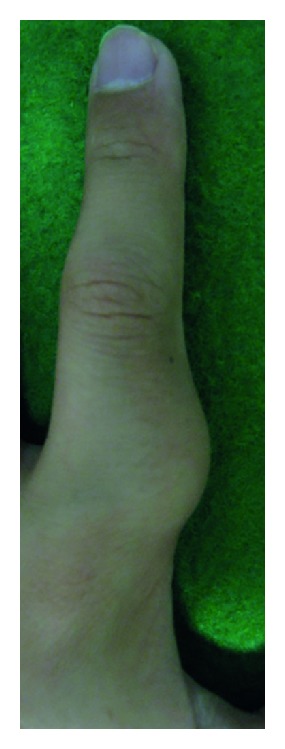
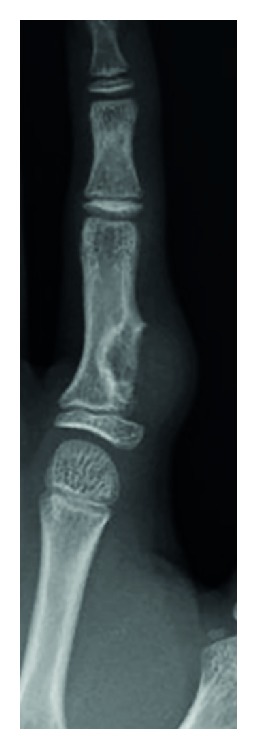
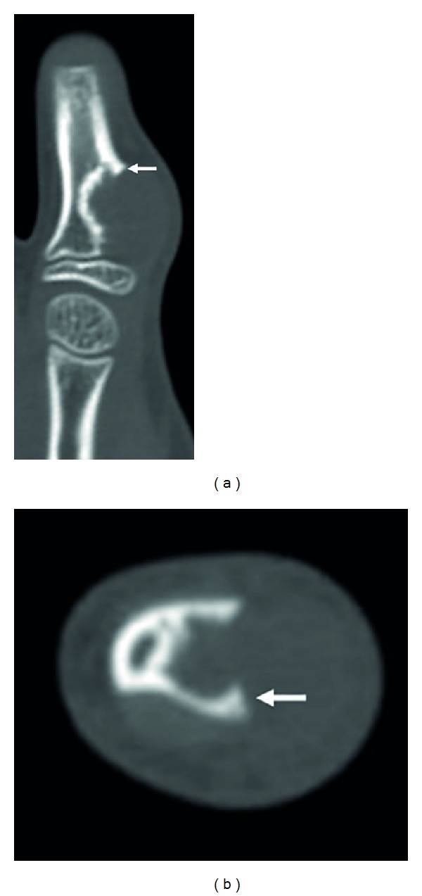
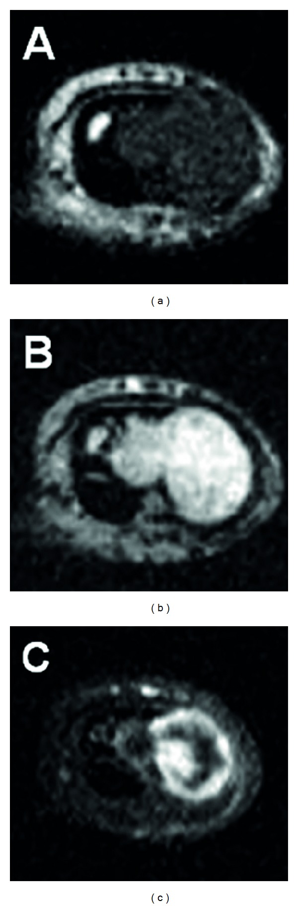
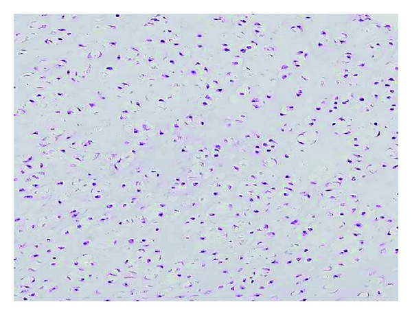
Periosteal chondroma is a rare benign hyaline cartilage neoplasm that occurs most commonly in the metaphases of long tubular bones. We present a unique case of periosteal chondroma arising in the proximal phalanx of the left index finger in a 12-year-old boy. Physical examination revealed a slightly protuberant, subcutaneous mass. Plain radiographs and computed tomography scans showed a periosteal lesion producing saucerization of the cortex and subjacent cortical sclerosis, without internal matrix calcification. On magnetic resonance imaging, the lesion exhibited intermediate signal intensity on T1-weighted images and high signal intensity on T2-weighted images. Contrast-enhanced fat-suppressed T1-weighted images demonstrated peripheral and septal enhancement. The patient underwent a marginal excision with curettage of the underlying bone cortex. Histological examination confirmed the diagnosis of periosteal chondroma. There has been no evidence of local recurrence eight months after surgery. Periosteal chondroma can protrude into the subcutaneous soft tissue causing a palpable mass. Recognition of the typical radiological features can lead to an accurate diagnosis of this rare condition.
Figures
Similar articles
-
Periosteal chondroma of the distal tibia: Computed tomography and magnetic resonance imaging characteristics and correlation with histological findings.Mol Clin Oncol. 2015 May;3(3):677-681. doi: 10.3892/mco.2015.492. Epub 2015 Jan 22. Mol Clin Oncol. 2015. PMID: 26137286 Free PMC article.
-
Periosteal chondroma presenting as a subcutaneous mass in the thumb.Clin Imaging. 2001 Nov-Dec;25(6):432-4. doi: 10.1016/s0899-7071(01)00336-9. Clin Imaging. 2001. PMID: 11733159
-
Periosteal chondroma of the rib: A case report.World J Clin Cases. 2022 Aug 16;10(23):8330-8335. doi: 10.12998/wjcc.v10.i23.8330. World J Clin Cases. 2022. PMID: 36159509 Free PMC article.
-
Periosteal chondroma of the proximal humerus: a case report and review of the literature.J Med Assoc Thai. 2006 Nov;89(11):1970-5. J Med Assoc Thai. 2006. PMID: 17205883 Review.
-
Large chondroma of the dural convexity in a patient with Noonan's syndrome. Case report and review of the literature.Neurocirugia (Astur). 2007 Jun;18(3):241-6. Neurocirugia (Astur). 2007. PMID: 17622464 Review.
Cited by
-
Periosteal Chondroma of the Pelvis: An Uncommon Tumor in an Unusual Location.Cureus. 2021 Aug 13;13(8):e17163. doi: 10.7759/cureus.17163. eCollection 2021 Aug. Cureus. 2021. PMID: 34548974 Free PMC article.
-
Ultrasound features of periosteal abnormalities in children.J Med Ultrason (2001). 2025 Jun 12. doi: 10.1007/s10396-025-01553-0. Online ahead of print. J Med Ultrason (2001). 2025. PMID: 40504318 Review.
-
Periosteal chondroma of the distal tibia: Computed tomography and magnetic resonance imaging characteristics and correlation with histological findings.Mol Clin Oncol. 2015 May;3(3):677-681. doi: 10.3892/mco.2015.492. Epub 2015 Jan 22. Mol Clin Oncol. 2015. PMID: 26137286 Free PMC article.
-
Bizarre Parosteal Osteochondromatous Proliferation Revisited.In Vivo. 2025 Jul-Aug;39(4):1799-1809. doi: 10.21873/invivo.13981. In Vivo. 2025. PMID: 40578997 Free PMC article. Review.
-
Extra-articular tenosynovial chondromatosis of the right fifth digit in a 59-year-old man: A case report and literature review.J Radiol Case Rep. 2021 Aug 1;15(8):8-17. doi: 10.3941/jrcr.v15i8.4212. eCollection 2021 Aug. J Radiol Case Rep. 2021. PMID: 35586796 Free PMC article. Review.
References
-
- Fletcher CDM, Bridge JA, Hogendoorn PCW, Mertens F. WHO Classification of Tumours of Soft Tissue and Bone. Lyon, France: IARC Press; 2013. Chondromas: enchondroma, periosteal chondroma; pp. 252–254.
-
- Nojima T, Unni KK, McLeod RA, Pritchard DJ. Periosteal chondroma and periosteal chondrosarcoma. American Journal of Surgical Pathology. 1985;9(9):666–677. - PubMed
-
- Yamamoto T, Nagira K, Kurosaka M. Periosteal chondroma presenting as a subcutaneous mass in the thumb. Clinical Imaging. 2001;25(6):432–434. - PubMed
-
- Yildirim C, Unay K, Rodop O, Gamsizkan M. Periosteal chondroma that presented as a subcutaneous mass in the ring finger. Journal of Plastic Surgery and Hand Surgery. 2011;45(2):117–120. - PubMed
-
- Amary MF, Bacsi K, Maggiani F, et al. IDH1 and IDH2 mutations are frequent events in central chondrosarcoma and central and periosteal chondromas but not in other mesenchymal tumours. Journal of Pathology. 2011;224(3):334–343. - PubMed
LinkOut - more resources
Full Text Sources
Other Literature Sources
