Review
. 2014 Sep;24(9):515-23.
doi: 10.1016/j.tcb.2014.04.003. Epub 2014 Apr 27.

Diverse cellular and molecular modes of axon degeneration

Affiliations
Review

Diverse cellular and molecular modes of axon degeneration

Lukas J Neukomm et al. Trends Cell Biol. 2014 Sep.

Abstract

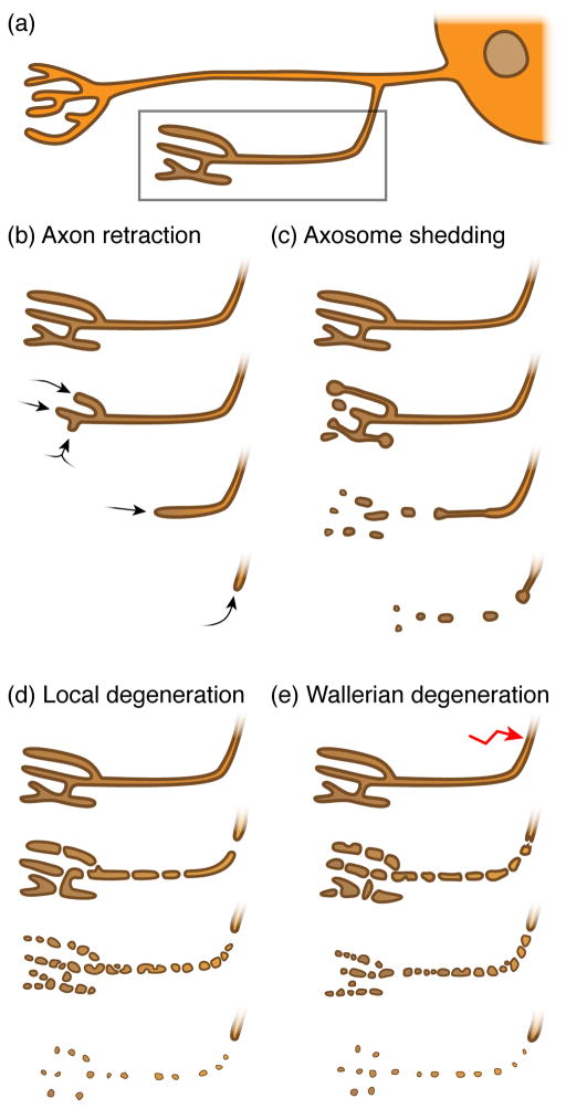
The elimination of large portions of axons is a widespread event in the developing and diseased nervous system. Subsets of axons are selectively destroyed to help fine-tune neural circuit connectivity during development. Axonal degeneration is also an early feature of nearly all neurodegenerative diseases, occurs after most neural injuries, and is a primary driver of functional impairment in patients. In this review we discuss the diversity of cellular mechanisms by which axons degenerate. Initial molecular characterization highlights some similarities in their execution but also argues that unique genetic programs modulate each mode of degeneration. Defining these pathways rigorously will provide new targets for therapeutic intervention after neural injury or in neurodegenerative disease.

Keywords: Wallerian degeneration; axon degeneration; axon retraction; axosome shedding; glia; pruning.


Figures

Figure 1
Figure 1. Morphologically distinct modes of axon degeneration
(a) Schematic neuron (soma including nucleus on the right) containing branched axons and two synapses (towards the left); dendrites are not shown. The synapse and its axon highlighted in the box will undergo various axon degenerative events over time, as shown in b – e. (b) During axon retraction, synapses & axons retract without the loss of axonal integrity. (c) While synapses and axons retract, they shed intact axoplasm-containing axosomes that are cleared by the surrounding glia in a process known as axosome shedding. (d) During local degeneration of axons, the synapse and its axon undergo catastrophic fragmentation, resulting in the removal of the axonal debris by surrounding glia. (e) During Wallerian degeneration, the distal axon separated from the soma undergoes catastrophic fragmentation. The surrounding glia then clears the resulting debris. Note that axon pruning and Wallerian degeneration share similar cellular qualities.
Figure 2
Figure 2. Axon retraction in the Dentate Gyrus
(a) In neonatal mice, granule neurons (orange) located in the Dentate Gyrus (DG) project their axons (so called mossy fibers) into both the main and the infrapyramidal tracts (MT and IPT, respectively) to terminate on CA3 pyramidal dendrites (dark grey). (b) After two months, the IPT is remodeled through stereotyped retraction (red arrow) to generate adult structures. (a’ & b’) At the molecular level, Sema3F expression correlates with the regressive time period: in neonatal mice, Sema3F is not detectable (− Sema3F), however, shortly after birth, Sema3F expression is upregulated (+ Sema3F). Sema3F binds to the Neuropilin-2 (Npn-2) receptor, which in turn releases β2-Chimaerin (β2Chn) to axonal membranes where it triggers the hydrolysis of GTP to GTP + Pi in Rac1, thereby leading to axon retraction. By contrast, growth cone repulsion occurs independently of β2Chn.
Figure 3
Figure 3. NGF-deprived local axon degeneration
(a) Nerve Growth Factor (NGF) withdrawal induces local degeneration of axons. Axon degeneration can be monitored in vitro upon local NGF withdrawal in neurons of the PNS using Campenot chambers [29], or in neurons of the CNS using microfluidic chambers [30]. (b) Molecular pathways mediating local axonal degeneration. Trophic factor deprivation leads to the activation of tumor necrosis factor (TNF) receptor family members such as death receptor 6 (DR6) or p75 neurotrophin receptor (p75NTR, also known as NGFR). They trigger the activation of specific members of the apoptotic signaling cascade, which results in pruning of axons. Receptor signaling leads to the upregulation and release of pro-apoptotic Bax from mitochondria, which triggers the activation of initiator caspase 9 and effector caspases 3 and 6. Bclw prevents pruning by inhibiting Bax. The role of Cytochrome c release from mitochondria in local axon degeneration remains unclear.
Figure 4
Figure 4. Molecular pathways mediating Wallerian degeneration
(a) Upon injury, the distal – from the soma separated – axon will undergo Wallerian degeneration (WD), which results in catastrophic axon self-destruction and clearance of resulting axonal debris by surrounding glial cells. WD can be attenuated, either by loss-of-function mutations of genes required for WD (green) or by over-expression of genes that inhibit it (red). (b) Factors that mediate WD are extracellular Ca2+ ions, dSarm/Sarm1 and the E3 ubiquitin ligase Highwire/Phr1. Loss of those candidates result in defective WD. In contrast, genes that antagonize WD are the neomorphic WldS and dNmnat/Nmnat2. Over-expression of those genes will lead to the inhibition of WD.

References

    1. Raff MC, et al. Axonal self-destruction and neurodegeneration. Science. 2002;296:868–871. - PubMed
    1. Luo L, O’Leary DDM. Axon retraction and degeneration in development and disease. Annu Rev Neurosci. 2005;28:127–156. - PubMed
    1. Medana IM, Esiri MM. Axonal damage: a key predictor of outcome in human CNS diseases. Brain. 2003;126:515–530. - PubMed
    1. Burke RE, O’Malley K. Axon degeneration in Parkinson’s disease. Exp Neurol. 2012 doi: 10.1016/j.expneurol.2012.01.011. - DOI - PMC - PubMed
    1. Johnson VE, et al. Axonal pathology in traumatic brain injury. Exp Neurol. 2013;246:35–43. - PMC - PubMed

Publication types