

Podocytes, signaling pathways, and vascular factors in diabetic kidney disease
- PMID: 24780459
- PMCID: PMC4075065
- DOI: 10.1053/j.ackd.2014.03.011
Podocytes, signaling pathways, and vascular factors in diabetic kidney disease
Abstract
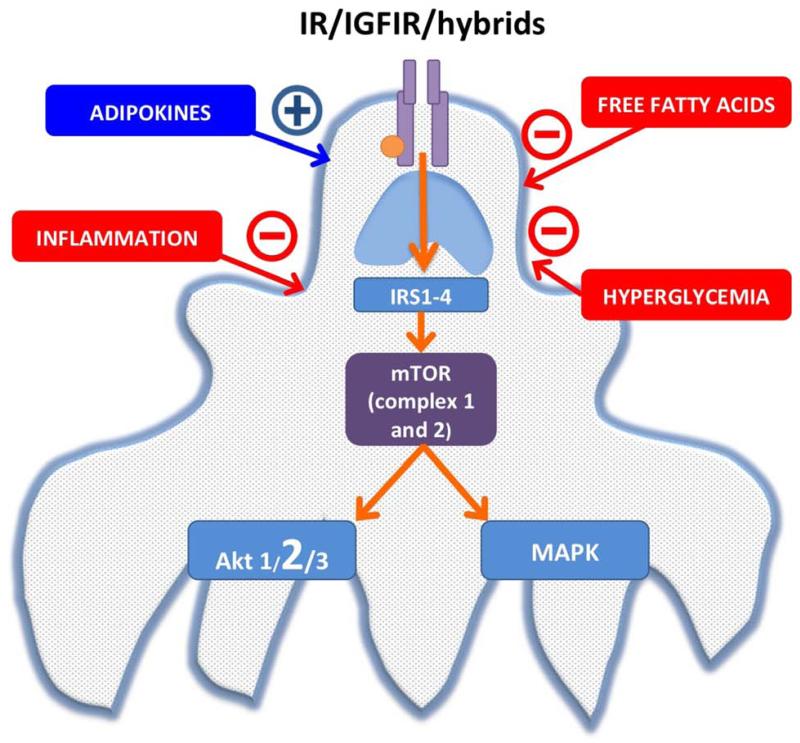
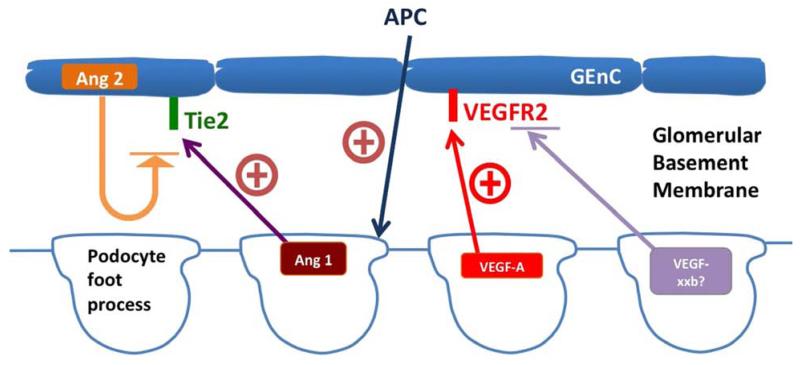
Alterations and injury to glomerular podocytes play a key role in the initiation and progression of diabetic kidney disease (DKD). Multiple factors in diabetes cause abnormalities in podocyte signaling that lead to podocyte foot process effacement, hypertrophy, detachment, loss, and death. Alterations in insulin action and mammalian target of rapamycin activation have been well documented to lead to pathology. Reduced insulin action directly leads to albuminuria, increased glomerular matrix accumulation, thickening of the glomerular basement membrane, podocyte apoptosis, and glomerulosclerosis. In addition, podocytes generate factors that alter signaling in other glomerular cells. Prominent among these is vascular endothelial growth factor-A, which maintains glomerular endothelium viability but causes endothelial cell pathology when generated at too high a level. Finally, circulating vascular factors (eg, activated protein C) have a profound effect on podocyte stability and survival. This cytoprotective factor is critical for podocyte health, and its deficiency promotes podocyte injury and apoptosis. Thus, the podocyte sits in the center of a network of paracrine and hormonal signaling systems that in health keep the podocyte adaptable and viable, but in diabetes they can lead to pathologic changes, detachment, and death.
Keywords: Diabetes; Glomerulus; Glucose; Insulin; Mammalian target of rapamycin.
Copyright © 2014 National Kidney Foundation, Inc. Published by Elsevier Inc. All rights reserved.
Figures
References
-
- Steffes MW, Schmidt D, McCrery R, Basgen JM. Glomerular cell number in normal subjects and in type 1 diabetic patients. Kidney Int. 2001 Jun;59(6):2104–2113. - PubMed
-
- Toyoda M, Najafian B, Kim Y, Caramori ML, Mauer M. Podocyte Detachment and Reduced Glomerular Capillary Endothelial Fenestration in Human Type 1 Diabetic Nephropathy. Diabetes. 2007 May 29; - PubMed
-
- Meyer TW, Bennett PH, Nelson RG. Podocyte number predicts long-term urinary albumin excretion in Pima Indians with Type II diabetes and microalbuminuria. Diabetologia. 1999 Nov;42(11):1341–1344. - PubMed
-
- Wharram BL, Goyal M, Wiggins JE, et al. Podocyte depletion causes glomerulosclerosis: diphtheria toxin-induced podocyte depletion in rats expressing human diphtheria toxin receptor transgene. J Am Soc Nephrol. 2005 Oct;16(10):2941–2952. - PubMed
Publication types
MeSH terms
Substances
Grants and funding
LinkOut - more resources
Full Text Sources
Other Literature Sources
Medical
Miscellaneous
