. 2014 Jul 15;391(2):147-57.
doi: 10.1016/j.ydbio.2014.04.015. Epub 2014 Apr 26.

Delayed transition to new cell fates during cellular reprogramming

Affiliations

Delayed transition to new cell fates during cellular reprogramming

Xianrui Cheng et al. Dev Biol. .

Abstract

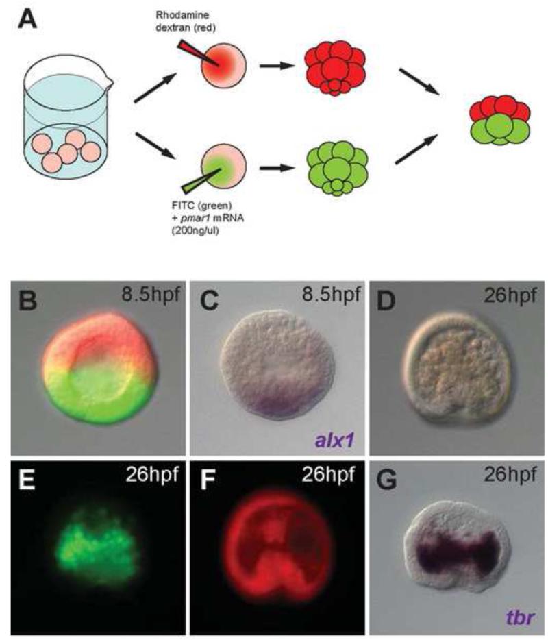
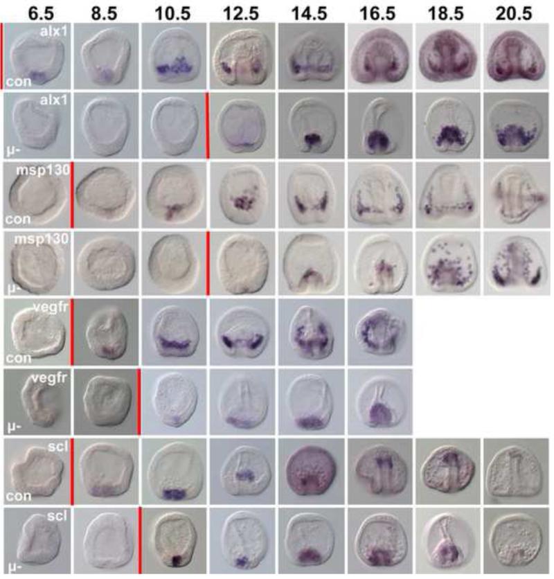
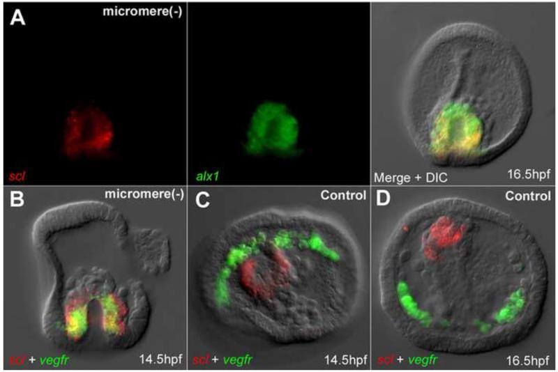
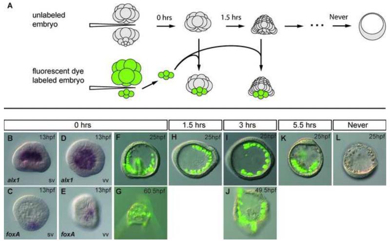
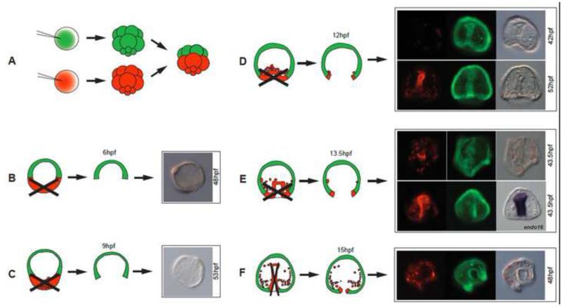
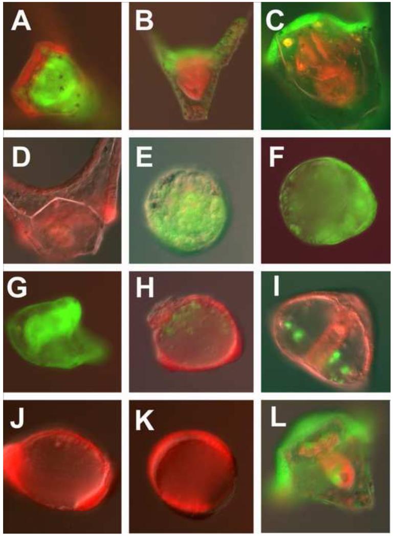
In many embryos specification toward one cell fate can be diverted to a different cell fate through a reprogramming process. Understanding how that process works will reveal insights into the developmental regulatory logic that emerged from evolution. In the sea urchin embryo, cells at gastrulation were found to reprogram and replace missing cell types after surgical dissections of the embryo. Non-skeletogenic mesoderm (NSM) cells reprogrammed to replace missing skeletogenic mesoderm cells and animal caps reprogrammed to replace all endomesoderm. In both cases evidence of reprogramming onset was first observed at the early gastrula stage, even if the cells to be replaced were removed earlier in development. Once started however, the reprogramming occurred with compressed gene expression dynamics. The NSM did not require early contact with the skeletogenic cells to reprogram, but the animal cap cells gained the ability to reprogram early in gastrulation only after extended contact with the vegetal halves prior to that time. If the entire vegetal half was removed at early gastrula, the animal caps reprogrammed and replaced the vegetal half endomesoderm. If the animal caps carried morpholinos to either hox11/13b or foxA (endomesoderm specification genes), the isolated animal caps failed to reprogram. Together these data reveal that the emergence of a reprogramming capability occurs at early gastrulation in the sea urchin embryo and requires activation of early specification components of the target tissues.

Keywords: Cell fate; Cell fate specification; Differentiation; Gene regulatory network; Regulative development; Reprogramming; Sea urchin embryo.


Figures

Figure 1
Figure 1
Skeletogenic reprogramming in micromere (−) embryos occurs after a long delay. (A) (Left panel) Illustration of the experiment. At the 16-cell stage micromeres were removed (micromere(−), blue), or at the mesenchyme blastula stage PMCs were removed (PMC(−), red). (Right panel) Explanation of the qPCR plots. (B-H) Analysis of (B) pmar1, (C) alx1, (D) tbr, (E) alx1, late, (F) vegfr, (G) msp130, early, and (H) msp130 late (H). Horizontal axes give hours post fertilization (hpf) until morphogenesis: MB, mesenchyme blastula; EG, early gastrula; MG, mid-gastrula; LG, late gastrula; PR, prism; PL, pluteus. In each plot, the normalization standard sample is indicated by a * sign, and has the relative expression value of 1.
Figure 2
Figure 2
(A) Experimental procedure for obtaining micromere(−) embryos overexpressing pmar1 in macromeres. (B) A micromere(−) embryo overexpressing pmar1 in its macromeres, with the animal cap cells labeled by rhodamine dextran (red) and progeny of pmar1-injected macromeres labeled by FITC (green). (C) alx1 in situ hybridization for the same embryo in (B) indicating the activation of alx1 in half of the embryo. (D-G) A sibling embryo of (B) developed to 26hpf. (D) The gastrulated embryo showed increased number of ingressed cells in the blastocoel. (E) The ingressed cells derived almost entirely from pmar1-injected macromere labeled with FITC (green) and started to aggregate on the sides of the archenteron. (F) Animal cap cells labeled with rhodamine dextran (red) contributed to the archenteron. (G) In situ hybridization of tbr for the same embryo in (D-F) showing that the FITC-labeled cells (green) were tbr expressing skeletogenic cells.
Figure 3
Figure 3
Reprogramming occurs with compressed temporal dynamics. WMISH analysis of alx1, msp130, vegfr, and scl expression in control embryos (Con) and in micromere(−) (μ-) embryos over time. Numbers at top are hours post fertilization (hpf). Red vertical bars mark the approximate time each row demonstrated expression of the gene in question. First expression of alx1 in controls actually occurs at 4.5hpf (see Fig. 1C). As indicated by the red bars, in controls there is a 4 hr interval between expression of alx1 and msp130 and vegfr. In the reprogramming NSM cells alx1 is seen at 12.5hpf while msp130 is seen within an hour of that, and vegfr actually precedes expression of alx1 suggesting temporal compression relative to the sequence in normal development.
Figure 4
Figure 4
During reprogramming PMC and NSM markers are co-expressed. (A) Expression of scl and alx1 in reprogramming NSMs at 16.5hpf. (B) Expression of alx1 and vegfr in reprogramming NSMs at 14.5hpf. (C-D) scl and vegfr expressed in separate lineages in control embryos at 14.5hpf (C), and 16.5hpf (D).
Figure 5
Figure 5
Animal cap cells lose an ability to regulate during early cleavage stages. (A) Schematic of the experiment. Unlabeled animal cap cells isolated from 32-cell stage embryos were cultured for 0, 1.5, 3, or 5.5hrs before receiving freshly isolated micromere quartets from fluorescently labeled 16-cell stage donor embryos. Numerals at the top of each arrow indicate the times elapsed before isolated animal halves received donor micromere quartets. (B-E) In situ images of embryos developed from an animal half receiving micromere quartet immediately after isolation, fixed at 13hpf. (B,D) in situ hybridization of alx1 in the chimera embryo. (C,E) in situ hybridization of foxA in the chimera embryo. (F-K) Embryos developed from an animal half receiving FITC labeled (green) micromere quartet. (F) micromeres received at 32-cell stage, imaged at 25hpf and (G) 60.5hpf; (H) micromeres received after 1.5hrs of isolated culture, imaged at 25hpf; (I) micromeres received after 3hrs of isolated culture, at 25hpf and (J) 49.5hpf; (K) micromeres received after 5.5hrs and imaged at 25hpf. (L) A dauer blastula developed from an isolated animal half that never received micromeres. SV side view, VV ventral view.
Figure 6
Figure 6
Animal caps reprogram. (A) Embryos were produced with animal caps and vegetal halves labeled with different fluorescent dyes and recombined at the 32-cell stage. (B-F) After 6, 9, 12, 13.5 and 15hpf the red and green halves were separated. (B,C) Halves separated at 6 and 9hpf did not develop beyond dauer blastulae. (D-F) Animal halves isolated at 12, 13.5 or 15hpf, contributed to vegetal germ layers (7/11 at 12hpf; 3/4 at 13.5hpf; 4/9 at 15hpf in this experiment). In E, lower right panel, in situ for endo16 of the same embryo seen to its left.
Figure 7
Figure 7
Endoderm GRN necessary for reprogramming. Red/green half embryos were recombined at the 32-cell stage and imaged at 48 hpf. In each case the embryos were imaged in both the red and green channels and merged. (A). Control red animal cap embryo recombined with a green vegetal half embryo. (B). Green Hox11/13b knockdown in the animal cap recombined with a control red vegetal half embryo. (C). Green FoxA knockdown in the animal cap recombined with control red vegetal half. Note: there is no mouth. (D). Control red animal cap after removal of green control vegetal half at EG. (E). Green Hox11/13b knockdown animal cap after removal of control red vegetal half at EG. (F). Green FoxA knockdown animal cap after removal of control red vegetal half at EG. (G). Control green animal cap after removal of control red vegetal half at EG. (H). Control red animal cap after removal of green Hox11/13b knockdown vegetal half at EG. (I). Control red animal cap after removal of green FoxA knockdown vegetal half at EG. (J). Red animal cap isolated at 32-cell stage and imaged at 48hpf. (K). Red animal cap recombined with green control vegetal half at 32-cell stage, isolated at HB, and imaged at 48hpf. (L). Green animal cap recombined with red vegetal half at 32-cell stage and half the red vegetal half removed at HB. HB, hatched blastula (7.5hpf); EG, early gastrula (13-14hpf).

References

    1. Amemiya S. Complete regulation of development throughout metamorphosis of sea urchin embryos devoid of macromeres. Dev Growth Differ. 1996;38:465–476. - PubMed
    1. Cherry AB, Daley GQ. Reprogramming cellular identity for regenerative medicine. Cell. 2012;148:1110–1122. - PMC - PubMed
    1. Croce JC, McClay DR. Dynamics of Delta/Notch signaling on endomesoderm segregation in the sea urchin embryo. Development. 2010;137:83–91. - PMC - PubMed
    1. Davidson EH. Academic; Burlington, MA; San Diego: 2006. The regulatory genome: gene regulatory networks in development and evolution.
    1. Davidson EH, Rast JP, Oliveri P, Ransick A, Calestani C, Yuh CH, Minokawa T, Amore G, Hinman V, Arenas-Mena C, Otim O, Brown CT, Livi CB, Lee PY, Revilla R, Rust AG, Pan Z, Schilstra MJ, Clarke PJ, Arnone MI, Rowen L, Cameron RA, McClay DR, Hood L, Bolouri H. A genomic regulatory network for development. Science. 2002;295:1669–1678. - PubMed

Publication types

LinkOut - more resources