Review
. 2014 Apr 28;29(2):139-45.
doi: 10.1016/j.devcel.2014.04.007.

Keeping at arm's length during regeneration

Affiliations
Review

Keeping at arm's length during regeneration

Valerie A Tornini et al. Dev Cell. .

Abstract

Regeneration of a lost appendage in adult amphibians and fish is a remarkable feat of developmental patterning. Although the limb or fin may be years removed from its initial creation by an embryonic primordium, the blastema that emerges at the injury site fashions a close mimic of adult form. Central to understanding these events are revealing the cellular origins of new structures, how positional identity is maintained, and the determinants for completion. Each of these topics has been advanced recently, strengthening models for how complex tissue pattern is recalled in the adult context.


Figures

Figure 1
Figure 1. Two Salamander Species Differ in the Cellular Origin of Regenerated Limb Muscle
(A) The newt (Notophthalmus viridescens) regenerates muscle after limb amputation, at least in part through the fragmentation and proliferation of mononuclear cells (red) derived from existing myofibers. PAX7+ satellite cells appear to be an insignificant source of blastemal tissue. (B) Existing myofibers are a minor component of muscle regeneration in the axolotl (Ambystoma mexicanum). Instead, PAX7+ satellite cells (green) are abundant, broadly distributed in the limb blastema, and a prime candidate for the major source of new muscle. Concepts based on Sandoval-Guzmán et al., 2014.
Figure 2
Figure 2. Adult Zebrafish Pectoral Fins Maintain Region-Specific Profiles of Patterning Transcription Factors Throughout Life
(A) Top view of the zebrafish (Danio rerio). Each of the five fin types regenerates completely after injury. The pectoral fin is highlighted, with its orientation marked as A (anterior) and P (posterior). (B) (Left) White shading in bars represents higher expression levels of the respective genes along the anteroposterior (AP) axis of pectoral fins, with lower expression levels proportionately fading to black. Each ray along the AP axis maintains a unique signature of different levels of alx4a, lhx9a, id4, pax9, tbx2a, hoxc8a, hoxd11a, hoxd13a, and hand2 in osteoblasts, fibroblasts, and/or other cell types. (Right) Such expression signatures are represented by unique colorations for each ray in this image. These signatures are retained during injury-induced regeneration, making this regulation a strong candidate to contribute to memory of position and restoration of pattern (Nachtrab et al., 2013).
Figure 3
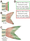
Figure 3. Calcineurin Is a Regulatory Target for Completion of Regeneration
(A–C) Model for how Calcineurin activity controls regeneration, based on concepts from Kujawski et al., 2014. Caudal fins are shown, which have a dorsal lobe (top) and a ventral lobe (bottom). (A) Following amputation, a regenerative growth program, represented in green, initiates. As part of this program, FK506 binding protein (FKBP) levels increase at 3 days post amputation (dpa) while Calcineurin phosphatase activity is limited. (B) As the regenerating fin reaches its final pattern, Calcineurin activity levels increase, which in turn helps to slow and terminate regenerative growth. The regions and signals in which regenerative growth has terminated are represented in red. (C) Under continuous treatment with FK506, Calcineurin activity remains inhibited, the regenerative growth program continues, and the duration of regenerative growth is extended. (D) Relative gene expression levels associated with regenerative growth (green) or termination and return to homeostasis (red) in (A–C).

Similar articles

Cited by

References

    1. Adams DS, Masi A, Levin M. H+ pump-dependent changes in membrane voltage are an early mechanism necessary and sufficient to induce Xenopus tail regeneration. Development. 2007;134:1323–1335. - PubMed
    1. Beane WS, Morokuma J, Lemire JM, Levin M. Bioelectric signaling regulates head and organ size during planarian regeneration. Development. 2013;140:313–322. - PMC - PubMed
    1. Blum N, Begemann G. Retinoic acid signaling controls the formation, proliferation and survival of the blastema during adult zebrafish fin regeneration. Development. 2012;139:107–116. - PubMed
    1. Broussonet PMA. Observations sur la regeneration de quelques parties du corps des Poissons. Hist de l’Acad Roy des Sciences 1786
    1. Chang HY, Chi JT, Dudoit S, Bondre C, van de Rijn M, Botstein D, Brown PO. Diversity, topographic differentiation, and positional memory in human fibroblasts. Proc Natl Acad Sci U S A. 2002;99:12877–12882. - PMC - PubMed

Publication types

LinkOut - more resources