Review
. 2014 Sep:126:51-60.
doi: 10.1016/j.exer.2014.01.010. Epub 2014 Apr 26.

Understanding photoreceptor outer segment phagocytosis: use and utility of RPE cells in culture

Affiliations
Review

Understanding photoreceptor outer segment phagocytosis: use and utility of RPE cells in culture

Francesca Mazzoni et al. Exp Eye Res. 2014 Sep.

Abstract

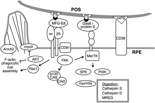
RPE cells are the most actively phagocytic cells in the human body. In the eye, RPE cells face rod and cone photoreceptor outer segments at all times but contribute to shedding and clearance phagocytosis of distal outer segment tips only once a day. Analysis of RPE phagocytosis in situ has succeeded in identifying key players of the RPE phagocytic mechanism. Phagocytic processes comprise three distinct phases, recognition/binding, internalization, and digestion, each of which is regulated separately by phagocytes. Studies of phagocytosis by RPE cells in culture allow specifically analyzing and manipulating these distinct phases to identify their molecular mechanisms. Here, we compare similarities and differences of primary, immortalized, and stem cell-derived RPE cells in culture to RPE cells in situ with respect to phagocytic function. We discuss in particular potential pitfalls of RPE cell culture phagocytosis assays. Finally, we point out considerations for phagocytosis assay development for future studies.

Keywords: engulfment; phagocytosis; photoreceptor outer segment renewal; retinal pigment epithelium; signaling.


Figures

Figure 1
Figure 1. Primary mouse RPE cells seeded at high and low density after purification significantly differ in cell shape and F-actin organization at confluence.
RPE cells were purified according to published procedures from PN8 wild-type mice (strain 129SvEms/J) and seeded at a density of yield of 2 eyes per well (A, lane 1/1, B and C) or diluted 8-fold (A, lane 1/8, D and E). Cells were analyzed after 6 days in culture. (A) Immunoblotting of protein content of one well of each cell population reveals that RPE cells seeded at either density express the tight junction protein ZO-1, the engulfment receptor MerTK, actin, and the visual cycle enzyme RPE65 (lanes as indicated). Cells seeded at lower density express lower levels of ZO-1, MerTK and actin. (B to E) Cells were fixed with 4% paraformaldehyde in PBS, permeabilized with 0.2% Triton × 100 and labeled with antibodies to ZO-1 (B and D, green) or Alexafluor488 conjugated phalloidin (C and E, green). Nuclei were counterstained with DAPI (shown in red). Maximal projections of image stacks acquired on a Leica TSP5 confocal microscopy system are shown. Scale bar: 20 μm.
Figure 2
Figure 2. Unpassaged primary mouse and rat RPE cells in culture and immortalized RPE-J cells are avid phagocytes.
RPE cells were purified according to published procedures from PN8 wild-type mice (A) or PN8 Long Evans rats (B) and seeded at a density of 2 eyes per well (A) and 1 eye per well (B). Immortalized rat RPE-J cells were seeded at 25% confluence (¼ dilution at split) on glass coverlips (C). All cells were challenged with 10 FITC-POS/cell 6 days after plating in DMEM supplemented with 0.2 ug/ml mouse MFG-E8 and 4% delipidated heat inactivated fetal bovine serum. Primary cells were challenged for 1.5 hours and RPE-J cells were challenged for 5 hours before fixation with ice-cold methanol. Nuclei were counterstained with DAPI. Images show representative maximal projections of image stacks of bound plus internalized FITC-POS (green) and RPE cell nuclei (red) obtained on a Leica TSP5 confocal microscopy system. For details of methods please see (Mao and Finnemann, 2013). Scale bars: 10 μm.
Figure 3
Figure 3. POS phagocytosis by RPE cells requires coordinated activities of numerous cell surface and cytosolic proteins.
The scheme illustrates our current knowledge on proteins involved in POS uptake gained from studies of both RPE phagocytosis in situ and in cell culture. Abbreviations: AnxA2, annexin A2; myoII, myosin II; myoVIIa, myosin VIIa; PI3K, PI3 kinases; SFK, Src family kinases. Adapted from (Mao and Finnemann, 2012). For details and references please see text.

Similar articles

Cited by

References

    1. Ahmado A, Carr AJ, Vugler AA, Semo M, Gias C, Lawrence JM, Chen LL, Chen FK, Turowski P, da Cruz L, Coffey PJ. Induction of differentiation by pyruvate and DMEM in the human retinal pigment epithelium cell line ARPE-19. Invest. Ophthalmol. Vis. Sci. 2011;52:7148–7159. - PubMed
    1. Albert DM, Tso MO, Rabson AS. In vitro growth of pure cultures of retinal pigment epithelium. Arch. Ophthalmol. 1972;88:63–69. - PubMed
    1. Besharse JC, Hollyfield JG, Rayborn ME. Photoreceptor outer segments: accelerated membrane renewal in rods after exposure to light. Science. 1977;196:536–538. - PubMed
    1. Bobu C, Hicks D. Regulation of retinal photoreceptor phagocytosis in a diurnal mammal by circadian clocks and ambient lighting. Invest. Ophthalmol. Vis. Sci. 2009;50:3495–3502. - PubMed
    1. Bok D, Hall MO. The role of the pigment epithelium in the etiology of inherited retinal dystrophy in the rat. J.Cell Biol. 1971;49:664–682. - PMC - PubMed

Publication types