. 2014 Jul;34(7):1233-42.
doi: 10.1038/jcbfm.2014.77. Epub 2014 Apr 30.

The contribution of ketone bodies to basal and activity-dependent neuronal oxidation in vivo

Affiliations

The contribution of ketone bodies to basal and activity-dependent neuronal oxidation in vivo

Golam M I Chowdhury et al. J Cereb Blood Flow Metab. 2014 Jul.

Abstract

The capacity of ketone bodies to replace glucose in support of neuronal function is unresolved. Here, we determined the contributions of glucose and ketone bodies to neocortical oxidative metabolism over a large range of brain activity in rats fasted 36 hours and infused intravenously with [2,4-(13)C₂]-D-β-hydroxybutyrate (BHB). Three animal groups and conditions were studied: awake ex vivo, pentobarbital-induced isoelectricity ex vivo, and halothane-anesthetized in vivo, the latter data reanalyzed from a recent study. Rates of neuronal acetyl-CoA oxidation from ketone bodies (V(acCoA-kbN)) and pyruvate (V(pdhN)), and the glutamate-glutamine cycle (V(cyc)) were determined by metabolic modeling of (13)C label trapped in major brain amino acid pools. V(acCoA-kbN) increased gradually with increasing activity, as compared with the steeper change in tricarboxylic acid (TCA) cycle rate (V(tcaN)), supporting a decreasing percentage of neuronal ketone oxidation: ∼100% (isoelectricity), 56% (halothane anesthesia), 36% (awake) with the BHB plasma levels achieved in our experiments (6 to 13 mM). In awake animals ketone oxidation reached saturation for blood levels >17 mM, accounting for 62% of neuronal substrate oxidation, the remainder (38%) provided by glucose. We conclude that ketone bodies present at sufficient concentration to saturate metabolism provides full support of basal (housekeeping) energy needs and up to approximately half of the activity-dependent oxidative needs of neurons.


Figures

Figure 1
Figure 1
Schematic depiction of the major metabolic pathways of 13C isotopic label flow from [2,4-13C2]BHB and the astroglial substrate, [2-13C]acetate. The continuous rate of metabolism of unlabeled glucose in neurons and astrocytes through pyruvate dehydrogenase, as represented by the rates, VpdhN or VpdhA, respectively, serves as constant dilution fluxes in these compartments. The cerebral metabolic rate for ketone body utilization (CMRkb) is related to the rate of acetyl-CoA oxidation from ketone bodies (VAcCoA-kb) as (CMRkb)/2=VAcCoA-kb. Ketone bodies and acetate are transported through membranes from blood to brain by monocarboxylic acid transporter (MCT) proteins. Subscripts on metabolite abbreviations (e.g., Ac2, αKG4, Glu4, GABA2, Gln4) refers to the initial (followed) carbon atom position labeled by 13C. AcAc, acetoacetate; Ac-CoA, acetyl-Coenzyme A; α-KG, α-ketoglutarate; BHB, β-hydroxybutyrate; GABA, gamma-aminobutyrate; Gln4, glutamine-C4; Glu4, glutamate-C4; lac, lactate; OAA, oxaloacetate; pyr, pyruvate; suc, succinate; VAc, rate of astroglial acetate oxidation; VAcCoA-kbN, VAcCoA-kbA, respective rates of neuronal and astroglial acetyl-CoA oxidation from ketone bodies; Vcyc, rate of glutamate-glutamine neurotransmitter cycle; VEff, efflux rate of glutamine; VGS, rate of glutamine synthesis; VPC, rate of pyruvate carboxylation; VtcaN, VtcaA; respective rates of neuronal and glial TCA cycles.
Figure 2
Figure 2
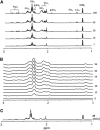
Stacked plots of 1H-[13C] difference spectra depicting cortical 13C metabolite labeling from the infused [2,4-13C2]BHB measured ex vivo in awake rats (A), in vivo in a halothane-anesthetized rat (B), and ex vivo during pentobarbital-induced isoelectricity (C). Intensities were scaled to the total creatine signal. Ala3, alanine-C3; Asp3, aspartate-C3; BHB4, β-hydroxybutyrate-C4; gaba2, gamma-aminobutyrate-C2; gaba3, gamma-aminobutyrate-C3; Gln3, glutamine-C3; Gln4, glutamine-C4; Glu3, glutamate-C3; Glu4, glutamate-C4; Lac3, lactate-C3; p.p.m., parts-per-million; time (in minutes) appears to the right of spectra.
Figure 3
Figure 3
Time courses of 13C labeling of cortical glutamate (Glu) and glutamine (Gln) during [2,4-13C2]BHB infusion in awake rats measured ex vivo with best-fits (solid lines) of the metabolic model. (A) Glutamate-C4,C3 percentage 13C enrichment verses infusion time. (B) Glutamine-C4 percentage 13C enrichment versus infusion time. Data shown reflect fractional enrichments as measured, without prior normalization by plasma 13C-BHB enrichment. (C) Monte-Carlo uncertainty distributions for VAcCoA-kbN and VpdhN based on 1,000 simulations. (D) Sensitivity of VAcCoA-kbN and VpdhN to changes in the assumed rate of astroglial anaplerosis (VPC) of ⩽25% above or below the nominal value (0.17 μmol/g per minute). Within the probable range of VPC, effects on the neuronal rates were small (<3.5%). BHB, β-hydroxybutyrate; VAcCoA-kbN, rate of acetyl-CoA oxidation from ketone bodies in neurons; Vcyc, rate of glutamate-glutamine neurotransmitter cycle; VPC, rate of pyruvate carboxylation via pyruvate carboxylase; VpdhN, rate of pyruvate oxidation in neurons via pyruvate dehydrogenase; VtcaN, rate of TCA cycle in neurons.
Figure 4
Figure 4
Plots of neuronal BHB oxidation (VAcCoA-kbN) and total TCA cycle rate (VtcaN) against glutamate-glutamine cycle rate (Vcyc) of fasted and BHB-infused rats under different states of neural activity (pentobarbital (PB)-induced isoelectricity, ∼1% halothane, and awake conditions). For the PB-isoelectric group VAcCoA-kbN is at or near saturation (maximum), whereas halothane and awake rates reflect non-saturating brain levels of BHB. The gray square depicts the calculated value of VAcCoA-kbN in awake rats under saturating blood BHB levels (from Figure 5) and a linear extrapolation (gray line) between the awake and isoelectric average rates, and given by: VAcCoA-kbN=0.84*Vcyc+0.23. For the halothane group, with Vcyc=0.37 μmol/g per minute, the estimated value of VAcCoA-kbN for saturating blood BHB level would be ∼0.54 μmol/g per minute, which is ∼30% higher than experimentally determined at the blood BHB level of 6.6 mM. For comparison with the ketone body data, the best linear fit for [1-13C]glucose-infused rats (small diamonds and dashed line with 95% confidence interval (CI)) from Rothman et al is shown (VtcaN=1.80(±0.10)Vcyc+0.19(±0.03), adjusted r2=0.97, n=12). The substantial degree of overlap in VtcaN between the two conditions suggests that hyperketonemia per se did not alter VtcaN or its relationship with neural activity and that ketone bodies can fully support basal (non-signaling) metabolism under the study conditions. Error bars reflect inter-animal group s.d. (isoelectricity, n=4; and halothane anesthesia, n=6) or Monte-Carlo s.d. of 1,000 simulations (awake rats, n=16). BHB, β-hydroxybutyrate; VAcCoA-kbN, rate of acetyl-CoA oxidation from ketone bodies in neurons; Vcyc, rate of glutamate-glutamine neurotransmitter cycle; VtcaN, rate of TCA cycle in neurons.
Figure 5
Figure 5
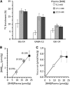
Dependence of brain amino acid 13C percentage enrichment and BHB level with increasing plasma BHB concentration in awake rat cortex. (A) 13C percentage enrichment of Glu-C4, GABA-C2, and Gln-C4 after a 7-minute infusion of [2,4-13C2]BHB of 1.5 (open square, n=4), 3.0 (gray square, n=5), or 4.5 (black square, n=5) mol/L resulting in the plasma concentrations shown. Brain amino acid 13C enrichments were corrected for 13C natural abundance by subtraction of 1.1% and divided by the respective plasma 13C-BHB enrichment. Values are reported as mean±s.d. (B) Relationship between brain (μmol/g) and plasma (mM) BHB concentration for the different groups plotted in (A) showing a near linear dependence with plasma concentration over the measured range of plasma BHB. The brain-to-plasma BHB ratio appears in parentheses above each group symbol with brain BHB concentration expressed as μmol/mL intracellular water assuming 0.77 mL/g ([BHB]brain in μmol/g × 1/0.77 mL/g). (C) Calculated rates of neuronal acetyl-CoA oxidation from ketone bodies (VAcCoA-kbN) at plasma (mM) BHB concentrations corresponding to the groups plotted in (A). VAcCoA-kbN for plasma BHB of 9.1 mM (0.40 μmol/minute per g) was taken from Table 3, whereas flux values for plasma BHB of 17 and 22 mM were calculated by multiplying the measured rate (0.40 μmol/min per g) by the respective percent increase (175%) in Glu-C4 enrichment reflected in (A). BHB, β-hydroxybutyrate; GABA, gamma-aminobutyrate; Gln, glutamine; Glu, glutamate; VAcCoA-kbN, rate of acetyl-CoA oxidation from ketone bodies in neurons.
Figure 6
Figure 6
Estimated fraction of total neuronal oxidation supported by ketone bodies and glucose for saturating levels of ketone bodies under pentobarbital (PB)-isoelectric and awake conditions. VAcCoA-kbN, rate of neuronal acetyl-CoA utilization from ketone bodies; VpdhN, neuronal pyruvate dehydrogenase flux (i.e., rate of acetyl-CoA utilization from glucose).

References

    1. Lopes-Cardozo M, Larsson OM, Schousboe A. Acetoacetate and glucose as lipid precursors and energy substrates in primary cultures of astrocytes and neurons from mouse cerebral cortex. J Neurochem. 1986;46:773–778. - PubMed
    1. Künnecke B, Cerdan S, Seelig J. Cerebral metabolism of [1,2-13C2]glucose and [U-13C4]3-hydroxybutyrate in rat brain as detected by 13C NMR spectroscopy. NMR Biomed. 1993;6:264–277. - PubMed
    1. Pan JW, de Graaf RA, Petersen KF, Shulman GI, Hetherington HP, Rothman DL. [2,4-13 C2]β-Hydroxybutyrate metabolism in human brain. J Cereb Blood Flow Metab. 2002;22:890–898. - PMC - PubMed
    1. Jiang L, Mason GF, RA Rothman DL, de Graaf RA, Behar KL. Cortical substrate oxidation during hyperketonemia in the fasted anesthetized rat in vivo. J Cereb Blood Flow Metab. 2011;31:2313–2323. - PMC - PubMed
    1. Sibson NR, Dhankhar A, Mason GF, Rothman DL, Behar KL, Shulman RG. Stoichiometric coupling of brain glucose metabolism and glutamatergic neuronal activity. Proc Natl Acad Sci USA. 1998;95:316–321. - PMC - PubMed

Publication types

LinkOut - more resources