Trichohyalin-like proteins have evolutionarily conserved roles in the morphogenesis of skin appendages
- PMID: 24780931
- PMCID: PMC4260798
- DOI: 10.1038/jid.2014.204
Trichohyalin-like proteins have evolutionarily conserved roles in the morphogenesis of skin appendages
Abstract
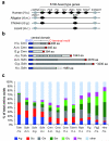
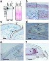
S100 fused-type proteins (SFTPs) such as filaggrin, trichohyalin, and cornulin are differentially expressed in cornifying keratinocytes of the epidermis and various skin appendages. To determine evolutionarily conserved, and thus presumably important, features of SFTPs, we characterized nonmammalian SFTPs and compared their amino acid sequences and expression patterns with those of mammalian SFTPs. We identified an ortholog of cornulin and a previously unknown SFTP, termed scaffoldin, in reptiles and birds, whereas filaggrin was confined to mammals. In contrast to mammalian SFTPs, both cornulin and scaffoldin of the chicken are expressed in the embryonic periderm. However, scaffoldin resembles mammalian trichohyalin with regard to its expression in the filiform papillae of the tongue and in the epithelium underneath the forming tips of the claws. Furthermore, scaffoldin is expressed in the epithelial sheath around growing feathers, reminiscent of trichohyalin expression in the inner root sheath of hair. The results of this study show that SFTP-positive epithelia function as scaffolds for the growth of diverse skin appendages such as claws, nails, hair, and feathers, indicating a common evolutionary origin.
Figures


Similar articles
-
The Trichohyalin-Like Protein Scaffoldin Is Expressed in the Multilayered Periderm during Development of Avian Beak and Egg Tooth.Genes (Basel). 2021 Feb 10;12(2):248. doi: 10.3390/genes12020248. Genes (Basel). 2021. PMID: 33578693 Free PMC article.
-
Cell differentiation in the embryonic periderm and in scaffolding epithelia of skin appendages.Dev Biol. 2024 Nov;515:60-66. doi: 10.1016/j.ydbio.2024.07.002. Epub 2024 Jul 2. Dev Biol. 2024. PMID: 38964706 Review.
-
Immunolocalization of scaffoldin, a trichohyalin-like protein, in the epidermis of the chicken embryo.Anat Rec (Hoboken). 2015 Feb;298(2):479-87. doi: 10.1002/ar.23039. Epub 2014 Sep 5. Anat Rec (Hoboken). 2015. PMID: 25142216
-
Existence of trichohyalin-keratohyalin hybrid granules: co-localization of two major intermediate filament-associated proteins in non-follicular epithelia.Differentiation. 1994 Nov;58(1):65-75. doi: 10.1046/j.1432-0436.1994.5810065.x. Differentiation. 1994. PMID: 7532602
-
S100 and S100 fused-type protein families in epidermal maturation with special focus on S100A3 in mammalian hair cuticles.Biochimie. 2011 Dec;93(12):2038-47. doi: 10.1016/j.biochi.2011.05.028. Epub 2011 Jun 1. Biochimie. 2011. PMID: 21664410 Review.
Cited by
-
VolcanoFinder: Genomic scans for adaptive introgression.PLoS Genet. 2020 Jun 18;16(6):e1008867. doi: 10.1371/journal.pgen.1008867. eCollection 2020 Jun. PLoS Genet. 2020. PMID: 32555579 Free PMC article.
-
Epidermal Differentiation Genes of the Common Wall Lizard Encode Proteins with Extremely Biased Amino Acid Contents.Genes (Basel). 2024 Aug 28;15(9):1136. doi: 10.3390/genes15091136. Genes (Basel). 2024. PMID: 39336727 Free PMC article.
-
Evolutionary diversification of epidermal barrier genes in amphibians.Sci Rep. 2022 Aug 10;12(1):13634. doi: 10.1038/s41598-022-18053-7. Sci Rep. 2022. PMID: 35948609 Free PMC article.
-
Convergent Evolution of Cysteine-Rich Keratins in Hard Skin Appendages of Terrestrial Vertebrates.Mol Biol Evol. 2020 Apr 1;37(4):982-993. doi: 10.1093/molbev/msz279. Mol Biol Evol. 2020. PMID: 31822906 Free PMC article.
-
Comparative genomics of sirenians reveals evolution of filaggrin and caspase-14 upon adaptation of the epidermis to aquatic life.Sci Rep. 2024 Apr 23;14(1):9278. doi: 10.1038/s41598-024-60099-2. Sci Rep. 2024. PMID: 38653760 Free PMC article.
References
-
- Alibardi L. Adaptation to the land: The skin of reptiles in comparison to that of amphibians and endotherm amniotes. J Exp Zool B Mol Dev Evol. 2003;298:12–41. - PubMed
-
- Alibardi L. Embryonic keratinization in vertebrates in relation to land colonization. Acta Zool. 2009;90:1–17.
-
- Barresi C, Stremnitzer C, Mlitz V, et al. Increased sensitivity of histidinemic mice to ultraviolet B radiation suggests a crucial role of endogenous urocanic acid in photoprotection. J Invest Dermatol. 2011;131:188–194. - PubMed
Publication types
MeSH terms
Substances
Grants and funding
LinkOut - more resources
Full Text Sources
Other Literature Sources
Molecular Biology Databases

