. 2014 Apr 29;9(4):e96400.
doi: 10.1371/journal.pone.0096400. eCollection 2014.

Increased Na⁺/Ca²⁺ exchanger expression/activity constitutes a point of inflection in the progression to heart failure of hypertensive rats

Affiliations

Increased Na⁺/Ca²⁺ exchanger expression/activity constitutes a point of inflection in the progression to heart failure of hypertensive rats

Jesica S Rodriguez et al. PLoS One. .

Abstract

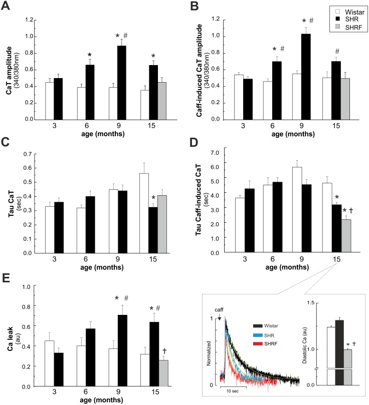
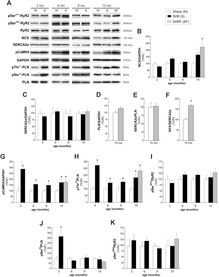
Spontaneously hypertensive rat (SHR) constitutes a genetic model widely used to study the natural evolution of hypertensive heart disease. Ca²⁺-handling alterations are known to occur in SHR. However, the putative modifications of Ca²⁺-handling proteins during the progression to heart failure (HF) are not well established. Moreover, the role of apoptosis in SHR is controversial. We investigated intracellular Ca²⁺, Ca²⁺-handling proteins and apoptosis in SHR vs. control Wistar rats (W) from 3 to 15 months (mo). Changes associated with the transition to HF (i.e. lung edema and decrease in midwall fractional shortening), occurred at 15 mo in 38% of SHR (SHRF). In SHRF, twitch and caffeine-induced Ca²⁺ transients, significantly decreased relative to 6/9 mo and 15 mo without HF signs. This decrease occurred in association with a decrease in the time constant of caffeine-Ca²⁺ transient decay and an increase in Na⁺/Ca²⁺ exchanger (NCX) abundance (p<0.05) with no changes in SERCA2a expression/activity. An increased Ca²⁺-calmodulin-kinase II activity, associated with an enhancement of apoptosis (TUNEL and Bax/Bcl2) was observed in SHR relative to W from 3 to 15 mo.

Conclusions: 1. Apoptosis is an early and persistent event that may contribute to hypertrophic remodeling but would not participate in the contractile impairment of SHRF. 2. The increase in NCX expression/activity, associated with an increase in Ca²⁺ efflux from the cell, constitutes a primary alteration of Ca²⁺-handling proteins in the evolution to HF. 3. No changes in SERCA2a expression/activity are observed when HF signs become evident.


Conflict of interest statement

Competing Interests: The authors have declared that no competing interests exist.

Figures

Figure 1
Figure 1. Progression from hypertrophy to heart failure in Wistar and SHR.
A) Systolic blood pressure (SBP). B) Left ventricular wall/tibial length ratio (LVW/TL). C) Echocardiographic left ventricular mass index (LVMI). D) Lung weight-to-tibial length ratio (LW/TL). E) Left ventricle midwall fractional shortening (MFS). All the above mentioned parameters were evaluated in normotensive (Wistar), hypertensive (SHR) and hypertensive with significant signs of contractile failure (SHRF), at 3, 6, 9, 12 and 15 months of age. Hypertension and hypertrophy were evident from early stages in SHR (A, B and C) while heart failure signs appeared at late stages (D and E). In 15 mo SHR, the evolution of the disease allowed to define two groups with significantly different contractile impairment. *p<0.05 with respect to W of the same age; †p<0.05 with respect to SHR at 15 mo; n≥8 animals per point.
Figure 2
Figure 2. Intracellular Ca2+ dynamics in isolated myocytes of Wistar and SHR.
A) Twitch Ca2+ transient (CaT) amplitude. B) Caffeine (Caff)-induced CaT amplitude. C) and D) Twitch and Caff-induced CaT decay parameters (tau). The inset in panel D) shows typical records of Caff-induced CaT (left) and diastolic Ca2+ (right) in W and SHR at 15 mo and in SHRF. E) Ca2+ leak from the SR. CaT amplitude and SR Ca2+ content (Caff-induced CaT) were higher in SHR at 6, 9 and 15 mo than in W of the same age. These increases were not observed in SHRF. At 15 mo there was a significant decrease in Tau of CaT and Caff-induced CaT in SHR relative to W. Tau of Caff-induced CaT in SHRF significantly diminished with respect to 15 mo SHR. At 9 and 15 mo there was an increase in SR Ca2+ leak in SHR vs. W, which did not occur in SHRF. *p<0.05 with respect to W of the same age; #p<0.05 with respect to SHR at 3 mo of age; †p<0.05 with respect to SHR 15 mo; n≥16 from at least 3 animals per group.
Figure 3
Figure 3. Activity of SERCA2a and relative contribution of NCX and SERCA2a to the Ca2+ transient constant.
A) The estimated SERCA2a activity (kSERCA2a) did not show any difference between SHR and W myocytes at all the time points studied. B) The relative contribution of NCX to the Ca2+ transient decay constant was significantly increased in SHRF. C) The relative contribution of SERCA2a to the Ca2+ transient decay constant was significantly decreased in SHRF. *p<0.05 with respect to W of the same age.
Figure 4
Figure 4. Time course of expression and phosphorylation of different proteins involved in intracellullar Ca2+ handling.
A) Representative immunoblots and average results of the expression and/or phosphorylation of B) NCX; C) SERCA2a; D) PLN; E) SERCA2a/PLN ratio; F) NCX/SERCA2a ratio; G) pCaMKII; H) pThr17-PLN; I) pSer2814-RyR2; J) pSer16-PLN; K) pSer2808-RyR2. The results are expressed as percentage of values obtained in W of the same age. Protein levels were normalized to the loading control glyceraldehyde 3-phosphate dehydrogenase (GAPDH). Phosphorylation of PLN and RyR2 was expressed as ratio between phosphorylated and non-phosphorylated forms of the proteins. While SERCA2a expression showed no differences at any time point studied, the expression of NCX was significantly higher only in SHRF. In this latter group, PLN expression and SERCA2a/PLN ratio did not change with respect to W, therefore the ratio NCX/SERCA2a was significantly enhanced. CaMKII and Thr17-PLN phosphorylations significantly increased from 3 mo in SHR with respect to W. PKA-dependent Ser16 phosphorylation of PLN increased at 3 mo and then decreased. Phosphorylation of Ser2808 and Ser2814 of RyR2 did not change at any age studied. *p<0.05 with respect to W of the same age; n≥4 animals per group.
Figure 5
Figure 5. Apoptosis in Wistar and SHR hearts.
A) Representative blots showing the expression of the proapototic (Bax) and antiapoptic (Bcl2) proteins in Wistar (W) and SHR (S) hearts at different ages. B) Quantitative analysis of protein expression, indicate increased Bax/Bcl2 ratio in SHR hearts at 6, 9 and 15 mo of age. (n≥4 animals per group). C) Typical example of TUNEL assay of 6 mo W and SHR and D) overall results of these experiments (n≥5 animals per group). Arrows indicate myocytes and arrow heads non-myocytes. Apoptosis is an early event in SHR hearts due to myocytes and non-myocytes death at 6 mo and to non-myocytes cells death at 15 mo. *p<0.05 with respect to W of the same age.

References

    1. Bers DM (2006) Altered cardiac myocyte Ca regulation in heart failure. Physiology (Bethesda) 21: 380–387. - PubMed
    1. Hadri L, Hajjar RJ (2011) Calcium cycling proteins and their association with heart failure. Clin Pharmacol Ther 90: 620–624. - PMC - PubMed
    1. Konstantinidis K, Whelan RS, Kitsis RN (2012) Mechanisms of cell death in heart disease. Arterioscler Thromb Vasc Biol 32: 1552–1562. - PMC - PubMed
    1. Wencker D, Chandra M, Nguyen K, Miao W, Garantziotis S, et al. (2003) A mechanistic role for cardiac myocyte apoptosis in heart failure. J Clin Invest 111: 1497–1504. - PMC - PubMed
    1. Olivetti G, Abbi R, Quaini F, Kajstura J, Cheng W, et al. (1997) Apoptosis in the failing human heart. N Engl J Med 336: 1131–1141. - PubMed

Publication types