. 2014 Apr;26(4):1544-1556.
doi: 10.1105/tpc.114.122986. Epub 2014 Apr 29.

ABORTED MICROSPORES Acts as a Master Regulator of Pollen Wall Formation in Arabidopsis

Affiliations

ABORTED MICROSPORES Acts as a Master Regulator of Pollen Wall Formation in Arabidopsis

Jie Xu et al. Plant Cell. 2014 Apr.

Abstract

Mature pollen is covered by durable cell walls, principally composed of sporopollenin, an evolutionary conserved, highly resilient, but not fully characterized, biopolymer of aliphatic and aromatic components. Here, we report that ABORTED MICROSPORES (AMS) acts as a master regulator coordinating pollen wall development and sporopollenin biosynthesis in Arabidopsis thaliana. Genome-wide coexpression analysis revealed 98 candidate genes with specific expression in the anther and 70 that showed reduced expression in ams. Among these 70 members, we showed that AMS can directly regulate 23 genes implicated in callose dissociation, fatty acids elongation, formation of phenolic compounds, and lipidic transport putatively involved in sporopollenin precursor synthesis. Consistently, ams mutants showed defective microspore release, a lack of sporopollenin deposition, and a dramatic reduction in total phenolic compounds and cutin monomers. The functional importance of the AMS pathway was further demonstrated by the observation of impaired pollen wall architecture in plant lines with reduced expression of several AMS targets: the abundant pollen coat protein extracellular lipases (EXL5 and EXL6), and CYP98A8 and CYP98A9, which are enzymes required for the production of phenolic precursors. These findings demonstrate the central role of AMS in coordinating sporopollenin biosynthesis and the secretion of materials for pollen wall patterning.


Figures

Figure 1.
Figure 1.
Coexpression Analysis of Candidate Genes Involved in Pollen Wall Formation. (A) Sixteen guide genes were selected from the 26 genes that have been demonstrated to be involved in pollen wall formation by forward genetics analysis. (B) Using coexpression analysis, 98 candidate genes were shown to be specifically expressed during pollen wall development. (C) About 43.8% of the genes (110/251) only matched one guide gene during the coexpression analysis, and the average frequency of each gene among 98 genes matching guide genes was more than 4. (D) Expression analysis of 11 selected genes by qRT-PCR analysis. Expression profiles of At1g71160 (KCS7), At3g52160 (KCS15), At5g49070 (KCS21), At1g75910 (EXL4), At1g75920 (EXL5), At1g75930 (EXL6), At1g06170 (bHLH089), At1g01280 (CYP703A2), At5g62080 (LTP), At2g16910 (AMS), and At4g14080 (MEE48/A6) as determined by qRT-PCR using total RNA from shoot (15 d), root (15 d), stem (15 d), leaf (15 d), seed, and anther samples. The relative amount of amplified product was normalized against At-ACT2 transcripts. Each value represents the average of two or more biologic replicate experiments in which each value is the mean of three separate qRT-PCR reactions (±se).
Figure 2.
Figure 2.
AMS Acts as a Key Transcriptional Regulator of Pollen Wall Formation in Arabidopsis. (A) and (B) Comparison of expression in male sterile mutants of 98 genes that may be involved in pollen wall formation. Mei, meiosis; PMI, pollen mitosis I; Bi, bicellular; PMII, pollen mitosis II; PCD, programmed cell death. (C) to (H) TEM analysis of pollen wall formation in the wild type (Columbia-0 [Col]) and ams. Stage 7 anthers ([C] and [D]), stage 8 anthers ([E] and [F]), and stage 10 anthers ([G] and [H]). Insets are a higher magnification of pollen exine showing tectum, bacula, and nexine. Msp, microspores; dMsp, degenerated microspores; pm, plasma membrane; PG, pollen grains. Bars = 100 μm for (C) to (H) and 1 μm for the insets. (I) to (N) TEM analysis of tapetal cells of the wild type (Col-0) and ams. Stage 7 anthers ([I] and [J]), stage 8 anthers ([K] and [L]), and stage 10 anthers ([M] and [N]) showing obvious accumulation of lipidic tapetosomes (arrow) and elaioplasts (arrow) in wild-type tapetal cells (M), and no detectable lipidic tapetosomes and elaioplasts in ams tapetal cells (N). T, tapetal cells; Nu, nucleus; Ela, elaioplasts; Ta, tapetosomes; Msp, microspores; dMsp, degenerated microspores. Bars = 5 μm. (O) to (T) Callose staining in wild-type (Col-0) and ams anthers. Stage 7 anthers ([O] and [P]), stage 9 anthers ([Q] and [R]), and stage 10 anthers ([S] and [T]) showing the delayed callose degradation in ams (arrow). Bars = 100 μm.
Figure 3.
Figure 3.
Metabolic Analysis of Wax, Cutin, Total Free Soluble Lipid, TPC, and TFC in Wild-Type and ams Buds. The wax, cutin, and total soluble lipid amounts expressed as dry weight (μg/mg) in the wild type and ams flower buds. The TPC and TFC amounts relative to standard substance content in each of 100 buds. Error bars indicate sd (n = 5). Some genes (shown in blue on pathway) had been reported as being involved in these metabolic processes; these genes are down in ams, which lead to changes of the corresponding compounds. [See online article for color version of this figure.]
Figure 4.
Figure 4.
Analysis of the Direct Target Genes of AMS. (A) qChIP-PCR analysis revealed the direct association of AMS with the promoters of 17 genes. DNA recovered from immunoprecipitation was used as templates for PCR using primer pairs (the double-headed arrows) spanning the putative AMS binding sequences in the upstream regions of candidate genes. Stars indicate the validation of the binding activity by AMS using EMSA. Data presented here represent the mean of five biological replicates. (B) EMSA of the binding of recombinant AMS onto the promoter fragments. DNA oligomers containing a consensus AMS recognition sequence found in the upstream regions of each of the candidate genes were labeled with digoxin and used as probes. (C) A network composed of AMS and correlated genes that are putatively involved in microspore compartmentalization in the tetrad, callose degeneration, lipid metabolism, and pollen exine formation and are direct targets of AMS, as determined using the AtGenExpress visualization tool on the GeneCAT website (http://genecat.mpg.de).
Figure 5.
Figure 5.
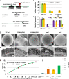
CYP98A8, CYP98A9, EXL5, and EXL6, Which Are Regulated by AMS, Are Required for Pollen Wall and Pollen Coat Development. (A) Schematic representation of the null cyp98A8/CYP98A9 RNAi double mutants derived from SALK-131366 T-DNA (green arrow) in CYP98A8 and RNAi (red arrow) in CYP98A9; SALK-057114 T-DNA (green arrow) in EXL5; and SALK-020241C T-DNA (green arrow) in EXL6. Green line represents the genome, black block represents the exons. (B) qRT-PCR of CYP98A8 and CYP98A9 expression in the wild type, ms null mutant, and null cyp98A8/CYP98A9 RNAi buds. Expression was normalized to ACTIN7 and presented relative to wild-type expression levels. Error bars represent sd of three biological replicates. (C) qRT-PCR analysis of the expression of EXL4, EXl5, and EXl6 in the wild-type, ams, exl5, and exl6 buds during anther development at stage 8. Expression was normalized to ACTIN7 and presented relative to wild-type expression levels with three biological replicates; error bars represent sd. (D) Analysis of the outer surface structure of pollen grains from the wild type, the null cyp98A8/CYP98A9 RNAi double mutant, and exl5 and exl6 mutants by scanning electron microscopy. Bars = 10 μm. (E) TEM analysis of the pollen wall from the wild type, the null cyp98A8/CYP98A9 RNAi double mutants, and the exl5 and exl6 mutants. Bars = 500 μm. (F) and (G) The standard curve of lipase activity (F) and lipase activity of pollen grains of ams, exl5, exl6, and the wild type (G). The values ​​were obtained under A420 wavelength measured, and assays were performed with three biological replicates.
Figure 6.
Figure 6.
Proposed Scheme Showing the Central Role of AMS in Pollen Wall Formation. AMS acts as a key transcriptional regulator that modulates the expression of genes associated with microspore compartmentalization and callose degeneration (A6 and QRT3), fatty acid/phenolic synthesis, elongation, metabolism and transport (KCS7, KCS15, KCS21, CYP703A2, CYP704B1, CYP86C3, CYP98A8, CYP98A9, PKSB/LAP5, TKPR1/DRL1, LTPs, and WBC27), and pollen coat formation (EXL4, EXL5, EXL6, GRP18, and GRP19). The micrographs refer to various developmental events of pollen wall in Arabidopsis. WBCs, White/Brown Complex subfamily. Bars = 30 μm. [See online article for color version of this figure.]

References

    1. Ahlers F., Thom I., Lambert J., Kuckuk R., Rolf W. (1999). 1H NMR analysis of sporopollenin from Typha angustifolia. Phytochemistry 50: 1095–1098
    1. Alonso J.M., et al. (2003). Genome-wide insertional mutagenesis of Arabidopsis thaliana. Science 301: 653–657 - PubMed
    1. Alves Ferreira M., de Almeida Engler J., Costa Miguens F., Van Montagu M., Engler G., de Oliveira D.E. (1997). Oleosin gene expression in Arabidopsis thaliana tapetum coincides with accumulation of lipids in plastids and cytoplasmic bodies. Plant Physiol. Biochem. 35: 729–739
    1. Ariizumi T., Toriyama K. (2011). Genetic regulation of sporopollenin synthesis and pollen exine development. Annu. Rev. Plant Biol. 62: 437–460 - PubMed
    1. Blackmore S., Wortley A.H., Skvarla J.J., Rowley J.R. (2007). Pollen wall development in flowering plants. New Phytol. 174: 483–498 - PubMed

LinkOut - more resources