. 2014;13(13):2029-37.
doi: 10.4161/cc.29018. Epub 2014 Apr 29.

Cockayne syndrome protein A is a transcription factor of RNA polymerase I and stimulates ribosomal biogenesis and growth

Affiliations

Cockayne syndrome protein A is a transcription factor of RNA polymerase I and stimulates ribosomal biogenesis and growth

Sylvia Koch et al. Cell Cycle. 2014.

Abstract

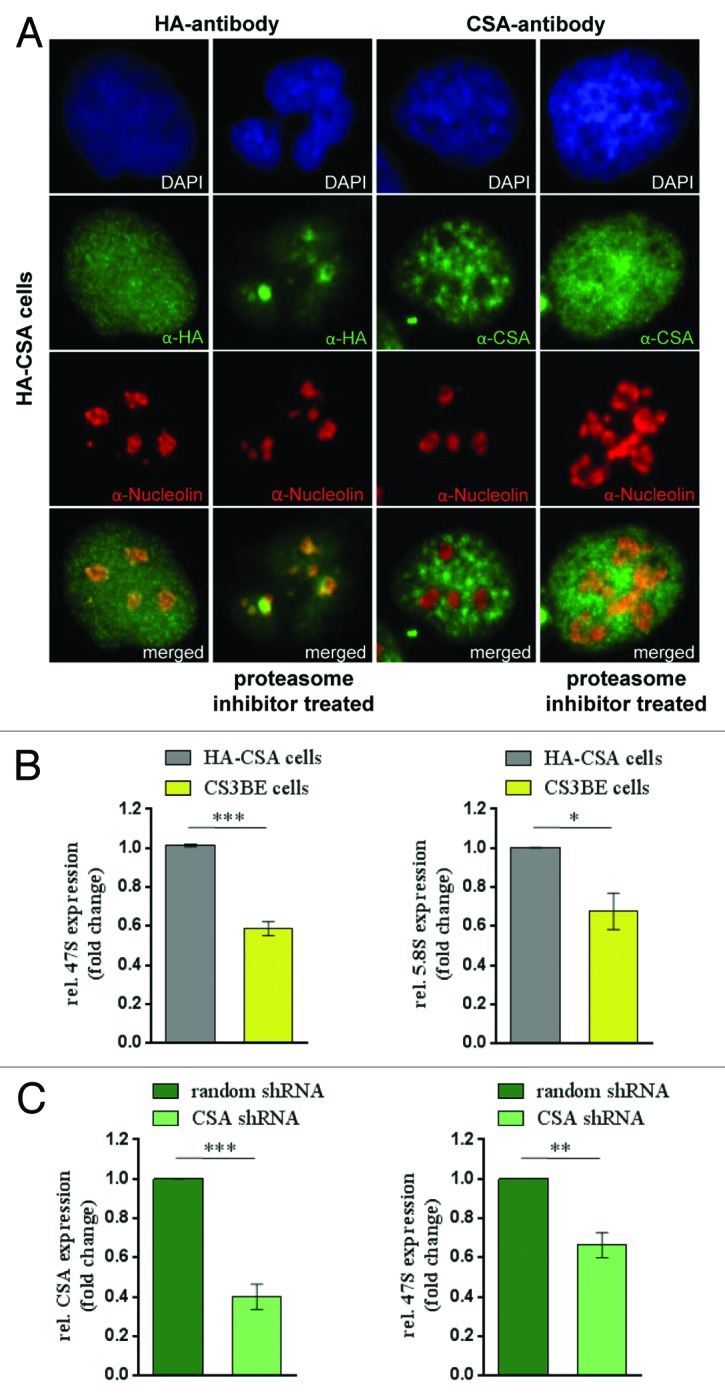
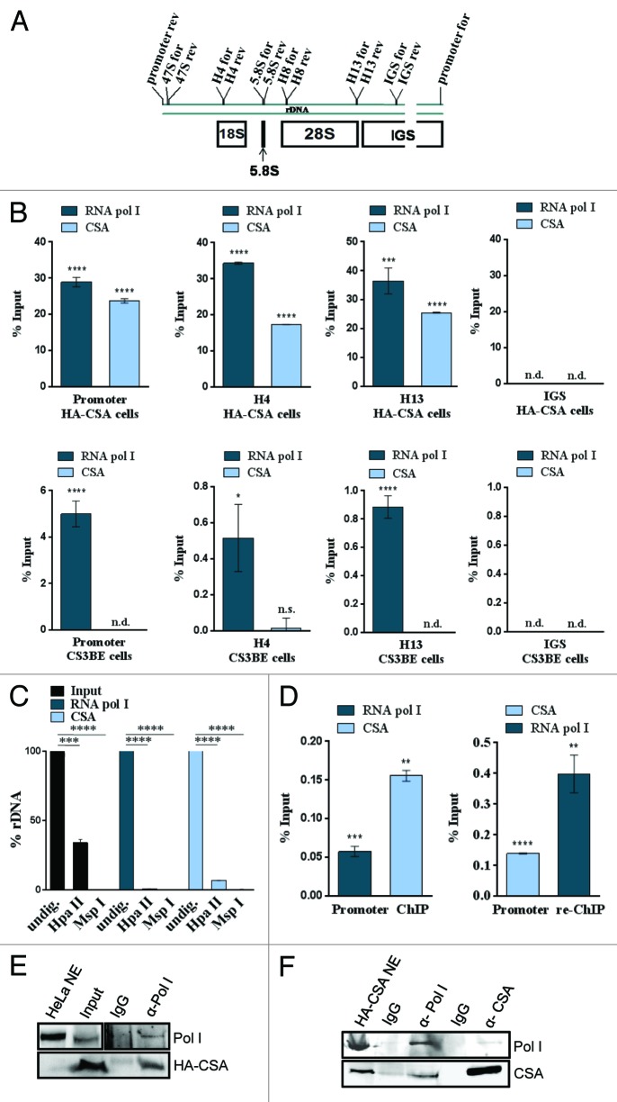
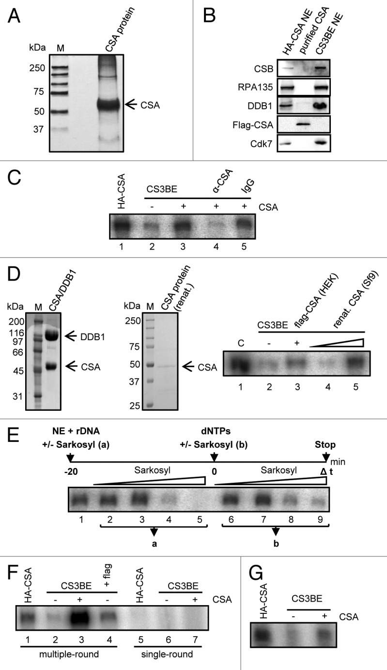
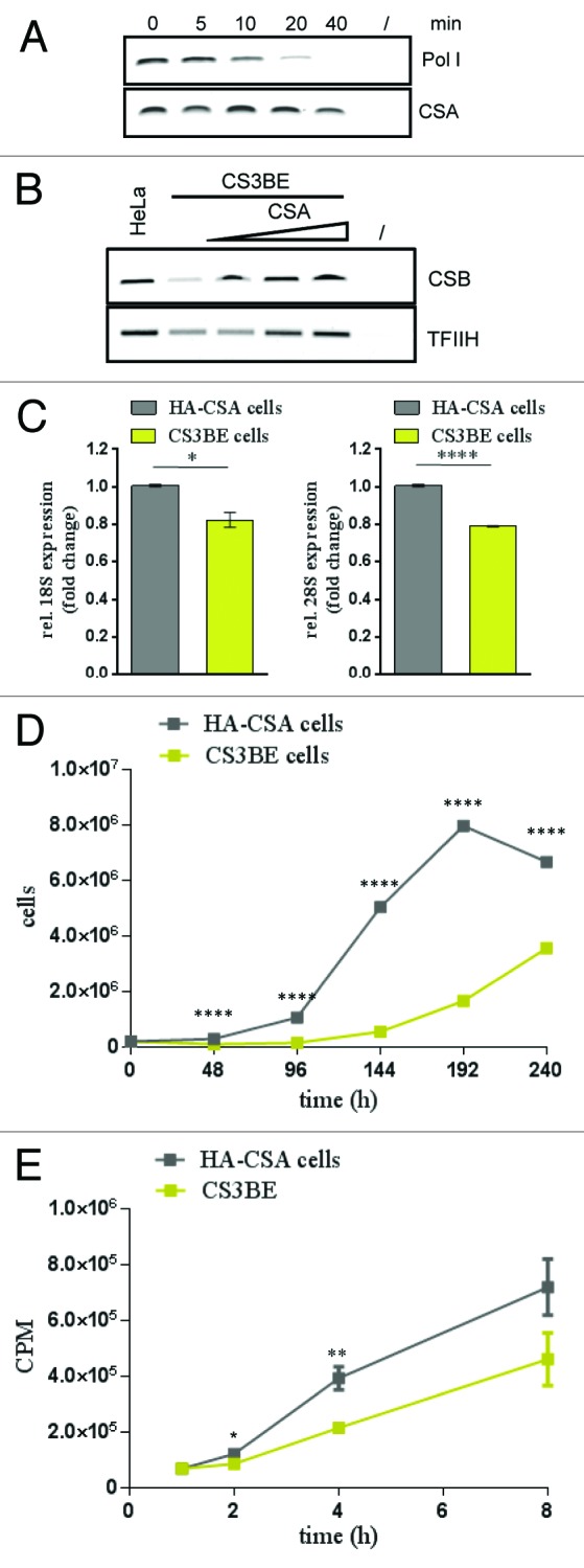
Mutations in the Cockayne syndrome A (CSA) protein account for 20% of Cockayne syndrome (CS) cases, a childhood disorder of premature aging and early death. Hitherto, CSA has exclusively been described as DNA repair factor of the transcription-coupled branch of nucleotide excision repair. Here we show a novel function of CSA as transcription factor of RNA polymerase I in the nucleolus. Knockdown of CSA reduces pre-rRNA synthesis by RNA polymerase I. CSA associates with RNA polymerase I and the active fraction of the rDNA and stimulates re-initiation of rDNA transcription by recruiting the Cockayne syndrome proteins TFIIH and CSB. Moreover, compared with CSA deficient parental CS cells, CSA transfected CS cells reveal significantly more rRNA with induced growth and enhanced global translation. A previously unknown global dysregulation of ribosomal biogenesis most likely contributes to the reduced growth and premature aging of CS patients.

Keywords: Cockayne syndrome; DNA repair; RNA polymerase I transcription; growth; premature aging; ribosomopathy.


Figures

None
Figure 1. CSA localizes to nucleoli and CSA mutation impairs rDNA transcription. (A) Immunofluorescence stainings with anti-HA and CSA antibody showing CSA in green and nucleolin in red in HA-CSA reconstituted CS3BE cells before and after proteasomal inhibition by MG-132. (B) QPCR analysis of the 47S precursor rRNA and the gene internal 5.8S/ internal transcribed spacer expression in CS3BE cells and CS3BE cells reconstituted with HA-tagged CSA. (C) Inhibition of CSA expression reduces rRNA transcription. QPCR detection of CSA expression and the 47S precursor rRNA expression after anti-CSA shRNA transfection and antibiotic selection in secondary fibroblasts. Values are normalized against RPL13 expression and the respective IgG controls. Values are mean ± s.e.m. of 3 independent experiments. (*P > 0.05; **P > 0.01; ***P > 0.001)
None
Figure 2. CSA associates with the active rDNA promoter and RNA polymerase I. (A) Schematic representation of a rDNA gene and the primers used in this study (IGS, intergenic spacer). (B) QPCR analysis of a representative ChIP experiment precipitated with the RNA polymerase I or HA-tag antibodies from chromatin of reconstituted (HA-CSA) or parental CS3BE cells. Values are normalized against input and IgG controls. (C) Methylation sensitive restriction analysis of ChIPed DNA reveals that CSA associates with the active, HpaII-digestable rDNA fraction like RNA polymerase I. (D) ChIP-re-ChIP experiment showing that CSA occupies the same molecules of rDNA as RNA polymerase I. Values are normalized against input and IgG controls. (E) ChIP–western experiment with the above indicated antibodies demonstrate that RNA polymerase I and CSA occupy the same rDNA molecules. (F) Co-immunoprecipitation with the above indicated antibodies and subsequent western blot analysis of 2 experiments with RNA polymerase I- and HA–CSA-specific antibodies. Pictures are representative of at least 3 independent experiments. Values are mean ± s.d. (*P > 0.05; **P > 0.01; ***P > 0.001; ****P > 0.0001)
None
Figure 3. CSA stimulates re-initiation of RNA polymerase I transcription. (A) Silver staining of purified CSA protein after SDS-PAGE. (B) Western blot analysis of purified CSA demonstrating that high-salt washed CSA is free of CSB, RNA polymerase I, DDB1, and TFIIH (cdk7). (C) In vitro RNA polymerase I transcription with the indicated nuclear extracts and addition of purified CSA and CSA-specific antibodies that specifically block stimulation. (D) Sf9-expressed CSA stimulates RNA polymerase I transcription. Coomassie staining of the Sf9 expressed DDB1/CSA complex and the renatured CSA. In vitro transcription reaction with renatured CSA protein. (E) Sarkosyl titration before (a) and after (b) initiation complex formation reveals the critical sarkosyl concentration for single-round transcription (compare lane 5 to 9). (F) Single-round transcription is not stimulated by CSA. Multiple but not single-round transcription is enhanced by addition of CSA. (G) Transcription with immobilized template showing that CSA enhances transcription after initiation complex formation. Bead-bound template was incubated with nuclear extract in the absence of CSA. After removal of the nuclear extract and washing of the preformed initiation complexes CSA was added and in vitro transcription performed. Pictures are representative of at least 3 independent experiments.
None
Figure 4. CSA recruits CSB and TFIIH to the rDNA promoter and loss of CSA retards ribosomal biogenesis and growth. (A) Template-immunoprecipitation experiment (13) showing the kinetics of CSA at the rDNA promoter after start of transcription. (B) Template immunoprecipitation experiments demonstrating CSB and TFIIH binding to the rDNA promoter after addition of CSA. (C) QPCR analysis of 18S and 28S rRNA amounts in cells lacking CSA (CS3BE) and CSA reconstituted cells. Values are normalized against RPL13 expression and the respective IgG controls and then normalized to HA-CSA cells. (D) Growth kinetics of CS3BE cells with or without CSA expression. (E) Metabolic labeling and translation kinetics of the indicated cells show that CSA stimulates protein biosynthesis. Pictures are representative of at least 3 independent experiments (A, B, D, and E) and values are mean ± s.e.m. of 3 independent experiments (C). (*P > 0.05; **P > 0.01; ***P > 0.001; ****P > 0.0001)

References

    1. Nance MA, Berry SA. Cockayne syndrome: review of 140 cases. Am J Med Genet. 1992;42:68–84. - PubMed
    1. Laugel V, Dalloz C, Durand M, Sauvanaud F, Kristensen U, Vincent MC, Pasquier L, Odent S, Cormier-Daire V, Gener B, et al. Mutation update for the CSB/ERCC6 and CSA/ERCC8 genes involved in Cockayne syndrome. Hum Mutat. 2010;31:113–26. doi: 10.1002/humu.21154. - DOI - PubMed
    1. Proietti-De-Santis L, Drané P, Egly JM. Cockayne syndrome B protein regulates the transcriptional program after UV irradiation. EMBO J. 2006;25:1915–23. doi: 10.1038/sj.emboj.7601071. - DOI - PMC - PubMed
    1. Itoh T, Ono T, Yamaizumi M. A new UV-sensitive syndrome not belonging to any complementation groups of xeroderma pigmentosum or Cockayne syndrome: siblings showing biochemical characteristics of Cockayne syndrome without typical clinical manifestations. Mutat Res. 1994;314:233–48. doi: 10.1016/0921-8777(94)90068-X. - DOI - PubMed
    1. Horibata K, Iwamoto Y, Kuraoka I, Jaspers NG, Kurimasa A, Oshimura M, Ichihashi M, Tanaka K. Complete absence of Cockayne syndrome group B gene product gives rise to UV-sensitive syndrome but not Cockayne syndrome. Proc Natl Acad Sci U S A. 2004;101:15410–5. doi: 10.1073/pnas.0404587101. - DOI - PMC - PubMed

Publication types

MeSH terms

LinkOut - more resources