. 2014 Apr 30:5:3673.
doi: 10.1038/ncomms4673.

G-protein stimulatory subunit alpha and Gq/11α G-proteins are both required to maintain quiescent stem-like chondrocytes

Affiliations

G-protein stimulatory subunit alpha and Gq/11α G-proteins are both required to maintain quiescent stem-like chondrocytes

Andrei S Chagin et al. Nat Commun. .

Abstract

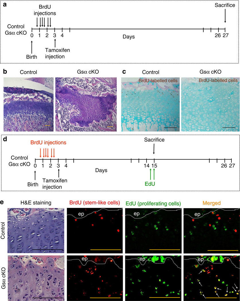
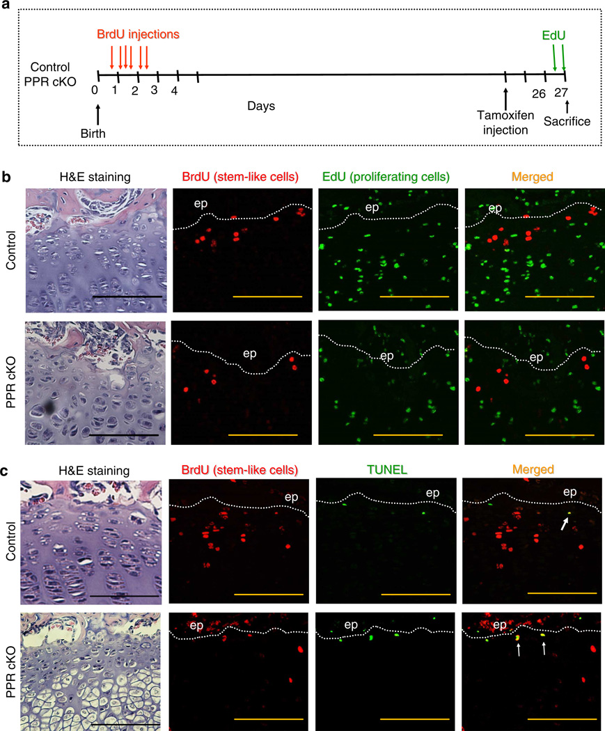
Round chondrocytes in the resting zone of the growth plate provide precursors for columnar chondrocytes and have stem-like properties. Here we demonstrate that these stem-like chondrocytes undergo apoptosis in the absence of the receptor (PPR) for parathyroid hormone-related protein. We examine the possible roles of heterotrimeric G-proteins activated by the PPR. Inactivation of the G-protein stimulatory α-subunit (G(s)α) leads to accelerated differentiation of columnar chondrocytes, as seen in the PPR knockout, but a remnant of growth cartilage remains, in contrast to disappearance of the growth cartilage in the PPR knockout. Stem-like chondrocytes lose their quiescence and proliferate upon G(s)α ablation. Inactivation of G(s)α in mice with a mutant PPR that cannot activate G proteins, Gq and G11, leads to a PPR knockout-like phenotype. Thus, G(s)α is the major mediator of the anti-differentiation action of the PPR, while activation of both G(s)α and Gq/11α is required for quiescence of stem-like chondrocytes.


Figures

Figure 1
Figure 1. Postnatal ablation of Gsα in chondrocytes leads to severe growth retardation and cartilage remnant formation
Representative pictures of control (Gsα fl/fl) and Gsα cKO (Col2-CreERt; Gsα fl/fl) (a) mice and (b) their long bones stained with Alizarin Red 1 month after tamoxifen administration. (c) Cre activity in the growth plate was monitored with a Rosa 26 reporter (Rosa). Control (Gsα fl/fl; Rosa/+) and Gsα cKO (Col2-CreERt; Gsα fl/fl; Rosa/+) mice were injected with tamoxifen, and x-gal staining was performed 5 days later. Sections were counterstained with Eosin. (d) Levels of Gsα mRNA were assayed 9 days after tamoxifen administration by in situ hybridization. Dashed lines depict the growth plate. ep, epiphyseal bone, (e) Time course of changes in the tibial growth plate upon ablation of Gsα or PTH receptor (PPR), which was performed utilizing an identical protocol and the same Col2-CreERt strain. Accelerated differentiation (day 5) followed by fusion of the growth plate (day 9, depicted by red arrowheads) was observed upon PPR ablation. Narrowing of the growth plate (day 9), followed by disorganization (day 13) and cartilage remnant formation (day 25) was observed upon Gsα ablation. In all experiments, tamoxifen was injected i.p. at 3 days of age. Scale bar, 100 µm (c-e).
Figure 2
Figure 2. Gsα ablation does not affect chondrocyte proliferation but accelerates differentiation and apoptosis
Tamoxifen was administrated at 3 days of age and growth plates were analysed 9 (a-d) and 13 (e-h) days later. Chondrocyte proliferation was not affected (a, pink cells, BrdU labelling for 2 hours), but post-mitotic flat cells were missed in Gsα cKO mice (depicted by yellow dotted line). Increase in the TUNEL-positive cells was observed at the chondro-osseous junction in Gsα cKO mice as compared with controls (b, green cells). At the same time, elevated numbers of cells expressing collagen type X mRNA (c) and Indian Hedgehog mRNA (d) were observed in Gsα cKO mice as compared with controls. (e) BrdU-positive chondrocytes (brown) were observed throughout the growth plate of Gsα cKO mice 13 days after gene ablation. Sections were counterstained with Eosin (pink) and Alcian Blue (blue). (f) TUNEL-positive cells (green) were detected mainly at the metaphyseal side of the growth plate but also at the epiphyseal side and in the middle of the growth plate 13 days after gene ablation. (g) Collagen type X mRNA (pink) was detected in the middle of the growth plate in Gsα cKO mice at this time point. (h) There was no increase in osteoclast activity in Gsα cKO mice as detected by TRAP staining. Dotted lines depict growth plates. f = flat cell layer; h = hypertrophic layer. Arrows show typical apoptotic chondrocytes. ep = epiphyseal bone. Scale bars, 100 µm.
Figure 3
Figure 3. Characterization of the cartilage remnant
Tamoxifen was injected at 3 days of age and growth plates were analysed 3 (a) or 1 month later (b-g). (a) The remnant remained in the bone 3 months after tamoxifen injection. (b) BrdU incorporation was detected in the growth plate of control (Gsα fl/fl) mice, but not in the remnant of Gsα cKO mice (BrdU injected 4 and 24 h before death). Sections were counterstained with Eosin (bone, pink) and Alcian Blue (cartilage, blue). (c) Collagen X mRNA was not detected in the remnant. (d) Some remnant cells were positive for Ihh mRNA, marker for prehypertrophic chondrocytes. (e) Gsα mRNA was not detected in the remnant of Gsα cKO mice, while prehypertrophic chondrocytes were positive in control mice. (f) β-galactosidase activity was detected in the remnant of Gsα cKO; Rosa reporter mice. X-gal staining was performed on non-fixed, non-decalcified frozen sections and counterstained with eosin. R=Rosa26. (g) Recombination of GNAS1 gene was detected in DNA isolated from the microdissected remnant (yellow arrowhead, g). The non-recombinant band in Gsα cKO mice is prominent, presumably due to contamination with Cre-negative surrounding tissue during microdissection. The upper band in the heterozygous control corresponds to the floxed allele and the lower band to the wild-type allele. Scale bars, 100 µm.
Figure 4
Figure 4. Further molecular characterization of the cartilage remnant
Tamoxifen was injected at 3 days of age and growth plates were analysed 1 month later. (a) Mef2C protein was detected immunohistochemically in prehypertrophic and hypertrophic chondrocytes in control mice; a few positive cells were also detected in the cartilage remnant of Gsα cKO mice. (b) phospho-SMAD1/5 was detected immunohistochemically in prehypertrophic and columnar chondrocytes in control mice but not in the remnant. (c) In control mice, Sox9 protein was detected throughout the growth plate. The most intense signal was found in prehypertrophic chondrocytes, whereas only a few cells were positive for Sox9 in the remnant. Scale bars, 100 µm.
Figure 5
Figure 5. Chondrocytes of reserve zone are recruited into proliferation in Gsα cKO mice
(a) Protocol for BrdU labelling of reserve zone cells (stem-like chondrocytes). (b) Representative images of the growth plates of control (Gsα fl/fl) and Gsα cKO (Col2-CreERt; Gsα fl/fl) mice shown in a,c. Sections are stained with H&E. (c) BrdU-detection revealed BrdU-positive stem-like chondrocytes in control, but not in Gsα cKO growth plates. Sections were counterstained with eosin (bone, pink) and Alcian Blue (cartilage, blue). (d) Modified protocol for BrdU labelling of stem-like chondrocytes aiming to detect stem-like cell proliferation. Mice were labelled with BrdU exactly as in a, but were killed earlier and were labelled with EdU 4 and 20 h before death. (e) Sections from control (Gsα fl/fl) and Gsα cKO (Col2-CreERt; Gsα fl/fl) mice treated as shown in d were stained with both BrdU and EdU. Double BrdU- and EdU-labelled cells (arrows) were observed in Gsα cKO mice but almost never in control mice. ep = epiphyseal bone. Scale bars, 100 µm.
Figure 6
Figure 6. Cells of the reserve zone are not recruited into the proliferative pool but undergo apoptosis in PPR cKO mice
(a) Scheme showing BrdU labelling of reserve zone cells (stem-like chondrocytes) and EdU labelling of proliferating chondrocytes in the growth plate of control (PPR fl/fl) and PPR cKO (Col2-CreERt; PPR fl/fl) mice. (b) Sections from control and PPR cKO mice treated as shown in a were stained for both BrdU and EdU. Double BrdU- and EdU-labelled cells were not observed in PPR cKO mice. (c) Sections from control and PPR cKO mice treated as shown in a were stained for both BrdU and simultaneously for apoptosis (TUNEL). Increased number of double TUNEL+BrdU-positive cells (arrows) was observed in PPR cKO mice as compared with control mice. Scale bars, 100 µm.
Figure 7
Figure 7. Cells of the reserve zone do not undergo apoptosis in Gsα cKO mice
(a) Scheme showing BrdU labelling of reserve zone cells (stem-like chondrocytes) and EdU labelling of proliferating chondrocytes in the growth plate of control (Gsα fl/fl) and Gsα cKO (Col2-CreERt; Gsα fl/fl) mice. (b) Sections from control and Gsα cKO mice shown in a were stained for both BrdU and for a marker of apoptosis (TUNEL). There were no differences in the small number of double TUNEL + BrdU-positive cells (arrows). Scale bars, 100µm.
Figure 8
Figure 8. Characterization of DSEL mice alone and in combination with Gsα KO
(a–d) DSEL mice9. No changes were observed in the growth plate height (a), BrdU incorporation (b), apoptosis (c) and BrdU-labelled stem-like cells co-visualized with TUNEL-positive cells (d, experimental details are the same as for Fig. 7a) between mice with one copy of mutated PTH receptor (DSEL mutation, D/+) and two copies of the mutated receptor (D/D). (e) In mice with inactivated Gsα in their growth plate, the DSEL mutation of two copies of the PTH receptor increased the number of cells positive for cleaved caspase-3 as compared with those in which only one copy of the PTH receptor was mutated. Tamoxifen was injected at 3 days of age and mice were analysed 9 days later. Scale bars, 100 µm.
Figure 9
Figure 9. Simultaneous ablation of Gsα from chondrocytes and Gq/11α signalling from the PPR
A gene (D) encoding a PTH/PTHrP receptor mutated so that it cannot activate Gq/11 G-proteins was introduced into Col2-CreERt; Gsα fl/fl mice. Tamoxifen was administrated at 3 days of age. Mice without Gsα signalling (Col2-CreERt; Gsα fl/fl) and with one or two copies of mutated PPR (D/+ or D/D) were compared. (a,b) One month after tamoxifen injection, fusion of the growth plate was observed in all tibias (a) in the mice lacking Gsα having mutated PPR, whereas small remnant can still be observed in some femurs (two out of eight animals analysed, b), genotypes as indicated. (c) Cessation of chondrocyte proliferation (assessed as BrdU incorporation 11 days after tamoxifen administration) and (d) elevation of ectopic apoptosis in the resting zone (assessed by TUNEL 9 days after tamoxifen administration, arrows). (e) Stem-like chondrocytes were labelled with BrdU as in Fig. 7a and stained for both BrdU and for apoptosis (TUNEL). Dotted lines depict growth plates. ep = epiphyseal bone. Scale bars, 100 µm.
Figure 10
Figure 10. Levels of cyclin D1 are regulated by both Gsα and Gq/11 signalling from PTH receptor
Levels of cyclin D1 were compared between mice with one copy of mutated PTH receptor (DSEL mutation, D/+) and two copies of the mutated receptor (D/D) in the presence or absence of Gsα. No changes were observed in the levels of cyclin D1 in the presence of intact Gsα signalling and impaired Gq/11 signalling (a,b). Inactivation of Gq/11 signalling in the absence of one allele of Gsα decreases levels of cyclin D1 (c,d) as well as inactivation of Gq/11 in the absence of both allels of Gsα (e-h). Tamoxifen was injected at 3 days of age and levels of cyclin D1 were analysed by immonohistochemistry 12 days later (a-f) or 15 days later (g,h). Scale bars, 100 µm.

References

    1. Kember NF, Lambert BE. Slowly cycling cells in growing bone. Cell Tissue Kinet. 1981;14:327–330. - PubMed
    1. Kember NF. Cell division in endochondral ossification. A study of cell proliferation in rat bones by the method of tritiated thymidine autoradiography. J. Bone Joint Surg. Br. 1960;42B:824–839. - PubMed
    1. Abad V, et al. The role of the resting zone in growth plate chondrogenesis. Endocrinology. 2002;143:1851–1857. - PubMed
    1. Mackie EJ, Tatarczuch L, Mirams M. The growth plate chondrocyte and endochondral ossification. J. Endocrinol. 2011;211:109–121. - PubMed
    1. Kronenberg HM. Developmental regulation of the growth plate. Nature. 2003;423:332–336. - PubMed

Publication types

MeSH terms

Substances

LinkOut - more resources