. 2014 Apr 29;55(7):4175-85.
doi: 10.1167/iovs.13-12856.

Uveal melanoma cell growth is inhibited by aminoimidazole carboxamide ribonucleotide (AICAR) partially through activation of AMP-dependent kinase

Affiliations

Uveal melanoma cell growth is inhibited by aminoimidazole carboxamide ribonucleotide (AICAR) partially through activation of AMP-dependent kinase

Ahmad Al-Moujahed et al. Invest Ophthalmol Vis Sci. .

Abstract

Purpose: To evaluate the effects and mechanism of aminoimidazole carboxamide ribonucleotide (AICAR), an AMP-dependent kinase (AMPK) activator, on the growth of uveal melanoma cell lines.

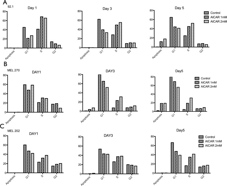
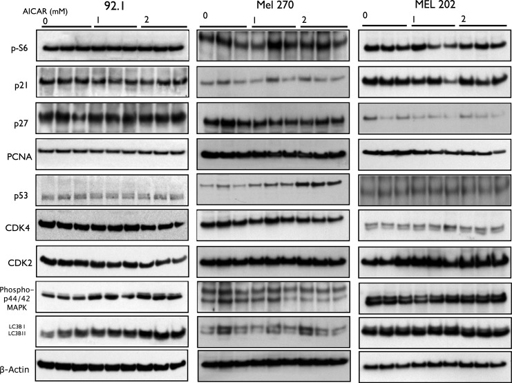
Methods: Four different cell lines were treated with AICAR (1-4 mM). Cell growth was assessed by 3-(4,5-dimethylthiazol-2-yl)-2,5-diphenyltetrazolium (MTT) assay. Cell cycle analysis was conducted by flow cytometry; additionally, expression of cell-cycle control proteins, cell growth transcription factors, and downstream effectors of AMPK were determined by RT-PCR and Western blot.

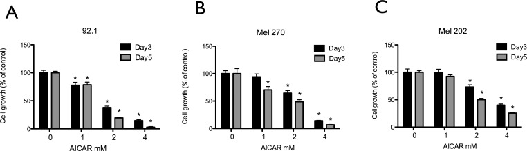
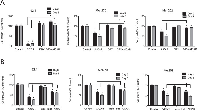
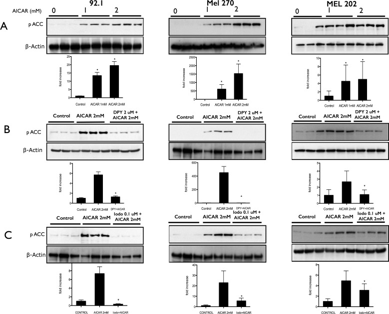
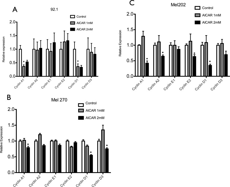
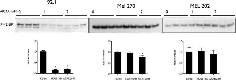
Results: Aminoimidazole carboxamide ribonucleotide inhibited cell growth, induced S-phase arrest, and led to AMPK activation. Aminoimidazole carboxamide ribonucleotide treatment was associated with inhibition of eukaryotic translation initiation factor 4E-BP1 phosphorylation, a marker of mammalian target of rapamycin (mTOR) pathway activity. Aminoimidazole carboxamide ribonucleotide treatment was also associated with downregulation of cyclins A and D, but had minimal effects on the phosphorylation of ribosomal protein S6 or levels of the macroautophagy marker LC3B. The effects of AICAR were abolished by treatment with dipyridamole, an adenosine transporter inhibitor that blocks the entry of AICAR into cells. Treatment with adenosine kinase inhibitor 5-iodotubericidin, which inhibits the conversion of AICAR to its 5'-phosphorylated ribotide 5-aminoimidazole-4-carboxamide-1-D-ribofuranosyl-5'-monophosphate (ZMP; the direct activator of AMPK), reversed most of the growth-inhibitory effects, indicating that some of AICAR's antiproliferative effects are mediated at least partially through AMPK activation.

Conclusions: Aminoimidazole carboxamide ribonucleotide inhibited uveal melanoma cell proliferation partially through activation of the AMPK pathway and downregulation of cyclins A1 and D1.

Keywords: AICAR; AMPK; mTOR; melanoma.


Figures

Figure 1
Figure 1
Aminoimidazole carboxamide ribonucleotide inhibits growth of human uveal melanoma cells. Uveal melanoma cell lines 92.1 (A), MEL 270 (B), and MEL 202 (C) were treated for 3 and 5 days with various concentrations of AICAR (1–4 mM), and cell viability was measured by MTT assay. Results are expressed as percentage of growth (%) relative to control values, defined as 100%. Data represent three independent experiments, each conducted with triplicate cultures. Significance (*) is assigned at P < 0.05.
Figure 2
Figure 2
Dipyridamole (DPY) and iodo effects on AICAR mediated uveal melanoma cell growth inhibition. Uveal melanoma cell lines 92.1, MEL 270, and MEL 202 were pretreated for 30 minutes with 2 μM DPY (A) or 0.1 μM iodo (B). Cells were then incubated for either 3 or 5 days without or with AICAR (2 mM). An MTT assay was performed, and results are expressed as percentage of growth (%) relative to control values, defined as 100%. Data represent three independent experiments, each conducted with triplicate cultures. Significance (*) is assigned at P < 0.05.
Figure 3
Figure 3
Aminoimidazole carboxamide ribonucleotide treatment of uveal melanoma cells is associated with activation of AMPK. (A) Western blot analysis of phosphorylated ACC (Ser-79) expression in 92.1, MEL 270, and MEL 202 cells that were treated with AICAR at a concentration of either 1 or 2 mM for 24 hours. (B) Western blot analysis of phosphorylated ACC expression in 92.1, MEL 270, and MEL 202 cells pretreated with DPY for 30 minutes before addition of AICAR at a concentration of 2 mM for 24 hours. (C) Western blot analysis of phosphorylated ACC expression in 92.1, MEL 270, and MEL 202 cells pretreated with iodo for 30 minutes before addition of AICAR at a concentration of 2 mM for 24 hours. Density values of phosphorylated ACC bands are graphically expressed relative to control. Multiple bands represent separate biological samples. Significance (*) is assigned at P < 0.05.
Figure 4
Figure 4
Aminoimidazole carboxamide ribonucleotide blocks cell cycle progression at S phase in human uveal melanoma cells. 92.1 (A), MEL 270 (B), and MEL 202 (C) uveal melanoma cells were treated with AICAR 1 and 2 mM for 1, 3, and 5 days. After overnight fixation, cells were suspended in PBS with RNase A and propidium iodide and acquired for DNA content by flow cytometry. All the data are graphically represented as percentage of cells in apoptosis, S phase, and G2/M phase. Data represent three independent experiments.
Figure 5
Figure 5
Aminoimidazole carboxamide ribonucleotide decreases the levels of different cyclins in uveal melanoma cells. 92.1 (A), MEL 270 (B), and MEL 202 (C) uveal melanoma cells were treated with AICAR at a concentration of either 1 or 2 mM for 24 hours. Quantitative RT-PCR analysis showed decrease of cyclins A1 and D1 in all cell lines,cyclin D3 in MEL 270 and cyclins A2 and E2 in MEL 202–treated cells in comparison with control cells. Significance (*) is assigned at P < 0.05.
Figure 6
Figure 6
Aminoimidazole carboxamide ribonucleotide does not affect levels of cell cycle progression regulators in uveal melanoma cells. Western blot analysis of phospho-S6, CDK inhibitor p21, CDK inhibitor p27, PCNA, p53 (except in Mel 270), CDK4, CDK2, phos-p44/p42 MAPK, and LC3B in 92.1, MEL 270, and MEL 202 cells treated with AICAR at a concentration of either 1 or 2 mM for 24 hours. Multiple bands represent separate biological samples.
Figure 7
Figure 7
Antiproliferative effect of AICAR on uveal melanoma cells is mediated via inhibition of 4E-BP1 phosphorylation in 92.1 and Mel 270, but not in Mel 202 cells. Western blot analysis of P-4E-BP1 in 92.1, Mel 720, and Mel 202 cells treated with AICAR at a concentration of either 1 or 2 mM for 24 hours. Density values of the bands are graphically expressed relative to control. Multiple bands represent separate biological samples. Significance (*) is assigned at P < 0.05.

References

    1. Landreville S, Agapova OA, Harbour JW. Emerging insights into the molecular pathogenesis of uveal melanoma. Future Oncol. 2008; 4: 629–636 - PMC - PubMed
    1. Bedikian AY. Metastatic uveal melanoma therapy: current options. Int Ophthalmol Clin. 2006; 46: 151–166 - PubMed
    1. Spagnolo F, Caltabiano G, Queirolo P. Uveal melanoma. Cancer Treat Rev. 2012; 38: 549–553 - PubMed
    1. Egan KMK, Seddon JMJ, Glynn RJR, Gragoudas ESE, Albert DMD. Epidemiologic aspects of uveal melanoma. Surv Ophthalmol. 1988; 32: 239–251 - PubMed
    1. Virgili G, Gatta G, Ciccolallo L, et al. Incidence of uveal melanoma in Europe. Ophthalmology. 2007; 114: 2309–2315, e2 - PubMed

Publication types

MeSH terms