Prevention of esophagopharyngeal reflux by augmenting the upper esophageal sphincter pressure barrier
- PMID: 24782387
- PMCID: PMC4332774
- DOI: 10.1002/lary.24735
Prevention of esophagopharyngeal reflux by augmenting the upper esophageal sphincter pressure barrier
Abstract
Objectives/hypothesis: Incompetence of the upper esophageal sphincter (UES) is fundamental to the occurrence of esophagopharyngeal reflux (EPR), and development of supraesophageal manifestations of reflux disease (SERD). However, therapeutic approaches to SERD have not been directed to strengthening of the UES barrier function. Our aims were to demonstrate that EPR events can be experimentally induced in SERD patients and not in healthy controls, and ascertain if these events can be prevented by application of a modest external cricoid pressure.
Study design: Individual case control study.
Methods: We studied 14 SERD patients (57 ± 13 years, 8 females) and 12 healthy controls (26 ± 3 years, 7 females) by concurrent intraesophageal slow infusion and pharyngoscopic and manometric technique without and with the application of a sustained predetermined cricoid pressure to induce, detect, and prevent EPR, respectively.
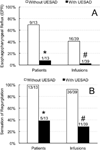
Results: Slow esophageal infusion (1 mL/s) of 60 mL of HCl resulted in a total of 16 objectively confirmed EPR events in none patients and none in healthy controls. All patients developed subjective sensation of regurgitation. Sustained cricoid pressure resulted in a significant UES pressure augmentation in all participants. During application of sustained cricoid pressure, slow intraesophageal infusion resulted in only one EPR event (P < .01).
Conclusions: Slow esophageal liquid infusion unmasks UES incompetence evidenced as the occurrence of EPR. Application of 20 to 30 mm Hg cricoid pressure significantly increases the UES intraluminal pressure and prevents pharyngeal reflux induced by esophageal slow liquid infusion. These techniques can be useful in diagnosis and management of UES incompetence in patients suffering from supraesophageal manifestations of reflux disease.
Keywords: Regurgitation; cricoid pressure; extraesophageal reflux disease; gastroesophageal reflux disease; laryngopharyngeal reflux; supraesophageal reflux disease.
© 2014 The American Laryngological, Rhinological and Otological Society, Inc.
Conflict of interest statement
The authors have no other funding, financial relationships, or conflicts of interest to disclose.
Figures


References
-
- Kamani T, Penney S, Mitra I, Pothula V. The prevalence of laryngopharyngeal reflux in the English population. Eur Arch Otorhinolaryngol. 2012;269:2219–2225. - PubMed
-
- Koufman JA. The otolaryngologic manifestations of gastroesophageal reflux disease (GERD): a clinical investigation of 225 patients using ambulatory 24-hour pH monitoring and an experimental investigation of the role of acid and pepsin in the development of laryngeal injury. Laryngoscope. 1991;101:1–78. - PubMed
-
- Sone M, Katayama N, Kato T, et al. Prevalence of laryngopharyngeal reflux symptoms: comparison between health checkup examinees and patients with otitis media. Otolaryngol Head Neck Surg. 2012;146:562–566. - PubMed
-
- Vakil N, van Zanten SV, Kahrilas P, Dent J, Jones R. The Montreal definition and classification of gastroesophageal reflux disease: a global evidence-based consensus. Am J Gastroenterol. 2006;101:1900–1920. quiz 1943. - PubMed
-
- Groome M, Cotton JP, Borland M, McLeod S, Johnston DA, Dillon JF. Prevalence of laryngopharyngeal reflux in a population with gastroesophageal reflux. Laryngoscope. 2007;117:1424–1428. - PubMed
Publication types
MeSH terms
Grants and funding
LinkOut - more resources
Full Text Sources
Other Literature Sources
Medical

