. 2014 Jun 12;123(24):3770-9.
doi: 10.1182/blood-2013-10-530964. Epub 2014 Apr 29.

CD28-mediated pro-survival signaling induces chemotherapeutic resistance in multiple myeloma

Affiliations

CD28-mediated pro-survival signaling induces chemotherapeutic resistance in multiple myeloma

Megan E Murray et al. Blood. .

Abstract

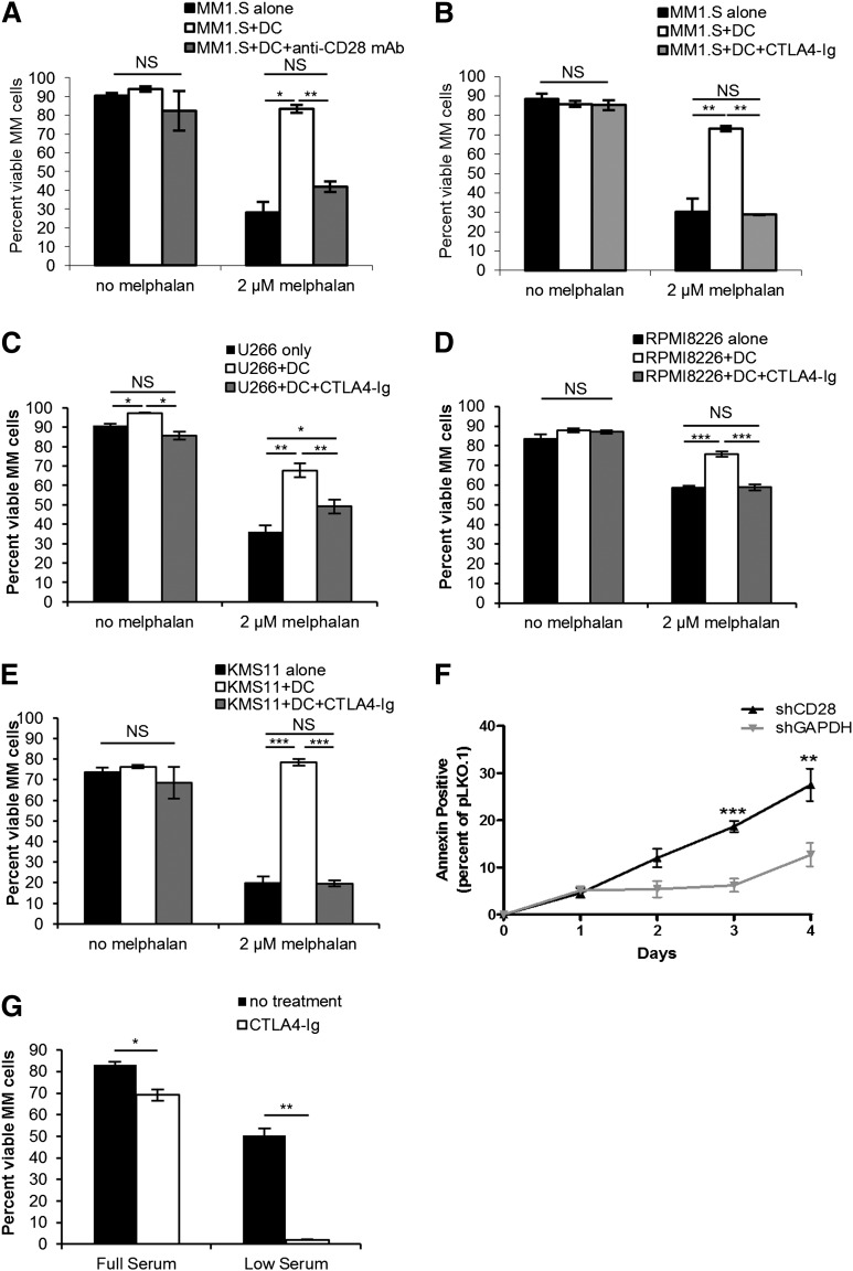
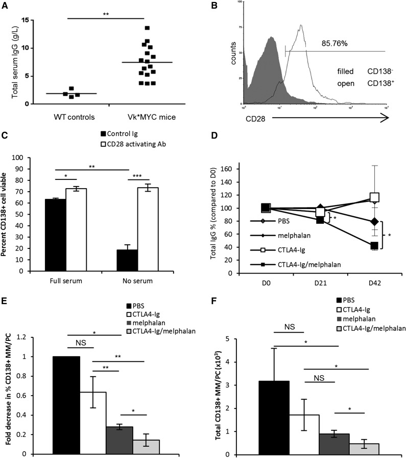
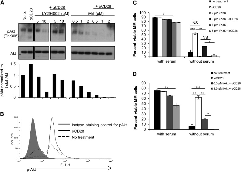
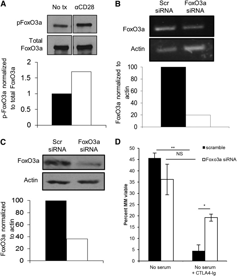
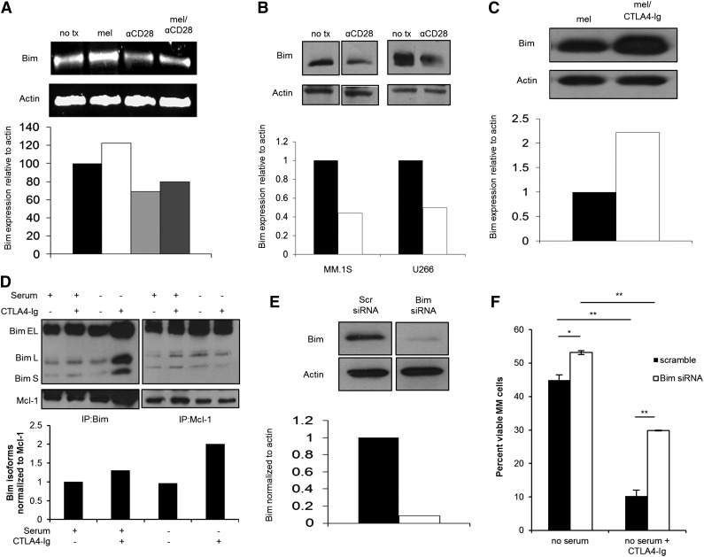
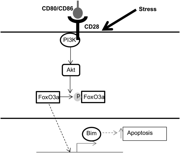
Chemotherapeutic resistance remains a significant hurdle in the treatment of multiple myeloma (MM) and is significantly mediated by interactions between MM cells and stromal cells of the bone marrow microenvironment. Despite the importance of these interactions, the specific molecules and downstream signaling components involved remain incompletely understood. We have previously shown that the prototypic T-cell costimulatory receptor CD28, which is also expressed on MM cells, is a key mediator of MM survival and apoptotic resistance. Crosslinking CD28 by agonistic antibodies or myeloid dendritic cells (DC; these express the CD28 ligands CD80/CD86) prevents apoptosis caused by chemotherapy or serum withdrawal. We now report that CD28 pro-survival signaling is dependent upon downstream activation of phosphatidyl-inositol 3-kinase/Akt, inactivation of the transcription factor FoxO3a, and decreased expression of the pro-apoptotic molecule Bim. Conversely, blocking the CD28-CD80/CD86 interaction between MM cells and DC in vitro abrogates the DC's ability to protect MM cells against chemotherapy-induced death. Consistent with these observations, in vivo blockade of CD28-CD80/CD86 in the Vk*MYC murine myeloma model sensitizes MM cells to chemotherapy and significantly reduces tumor burden. Taken together, our findings suggest that CD28 is an important mediator of MM survival during stress and can be targeted to overcome chemotherapy resistance.


Figures

Figure 1
Figure 1
CD28 signaling induced by DC or other MM cells prevents myeloma cell death. MM cells (MM.1S) were cultured alone or with DC at a 1:1 ratio ± 2 µM melphalan ± 50 µg/mL of the blocking αCD28 mAb CD28.6 (A) or ± 100 µg/mL CTLA4-Ig (B) or for 72 hours. Cell survival was analyzed by flow cytometry for Annexin V/7AAD staining and DC were gated out using CD11b. U266 (C), RPMI8226 (D), or KMS11 (E) myeloma cells were cultured as in (B) for 72 hours and survival was assessed by flow cytometry. (F) RPMI8226 myeloma cells were infected with lentiviral particles encoding shRNA specific for CD28, GAPDH, or empty vector (pLKO.1) and cultured for 4 days in full serum media. Survival was assessed by Annexin V staining. (G) MM.1S cells were cultured in either 10% serum or 0.2% (low) serum conditions for 5 days ± 100 µg/mL CTLA4-Ig. Fifty percent of the media was changed every other day to prevent nutrient depletion, and new CTLA4-Ig was added with every media change. Cell survival was analyzed by flow cytometry for Annexin V/7AAD staining. Data for panels A and B) are representative of 4 individual experiments; data for panels C-E are representative of 2 individual experiments. *P < .05, **P < .01, ***P < .005. NS, not significant.
Figure 2
Figure 2
CD28 signaling protects Vk*MYC murine myeloma cells in vitro and blockade sensitizes MM to melphalan in vivo. (A) Seventy-two-week-old Vk*MYC mice or their wild-type (WT) littermate controls were screened by ELISA for total serum IgG levels. (B) Whole bone marrow from Vk*MYC mice was isolated and the percent of myeloma cells was determined using a CD138+CD28+B220CD38MHCIICD3 phenotype. (C) Purified CD138+ cells from disease-bearing Vk*MYC mice were plated for 24 hours in media containing 10% fetal bovine serum (full serum) or no serum. Cells were cultured with either hamster Ig-coated beads (isotype control) or CD28-activating antibody-coated beads (clone PV1) at a ratio of 2 beads:1 cell. Cell viability was assessed by trypan blue exclusion. (D) Based on titers as in panel A, mice were randomized into 4 treatment groups (n = 3-4 mice/group): PBS, melphalan alone (2.0 mg/kg), CTLA4-Ig alone (100 μg/mouse), or melphalan plus CTLA4-Ig. Mice were treated intraperitoneally every third day for 42 days and serum samples were drawn weekly. Total serum IgG was determined by ELISA and was plotted as total IgG percent compared with day 0. (E) Percent MM/PC was determined using multiparametric flow for CD138+CD28+B220CD38MHCIICD3 cells, and fold decrease was calculated compared with the PBS group. (F) Total numbers of MM/PC were calculated by multiplying the percent MM/PC as determined in panel E by the total number of cells counted for each tissue. Data are representative of 2 separate experiments. *P < .05, **P < .01, ***P < .005.
Figure 3
Figure 3
CD28-mediated pro-survival signaling is dependent on PI3K and Akt. (A) MM.1S cells were cultured in full serum ±10 µg/mL CD28 activating mAb (CD28.2) and ± PI3K inhibitor LY294002 or ± iAkt II at the indicated doses. Cells were collected after 2 hours assessed by western blot. (B) MM.1S cells were cultured ± serum ± 10 µg/mL CD28.2 mAb. After 24 hours, cells were isolated, permeabilized, and stained intracellularly for phosphorylated Akt, which was assessed by flow cytometry. (C) MM.1S cells were cultured ± serum ±10 µg/mL CD28 activating mAb (CD28.2), and ± PI3K inhibitor LY294002 at the indicated doses. Cells were collected after 72 hours and viability was assessed via Annexin V and 7AAD staining by flow cytometry. (D) MM.1S cells were cultured ± serum ± 10 µg/mL αCD28.2 and ± Akt inhibitor (Akt inhibitor II) at the indicated doses. Cells were collected after 72 hours and viability was assessed via Annexin V and 7AAD staining by flow cytometry. Data for panels C-D) are representative of 3 independent experiments, and data for panels A-B are representative of 2 independent experiments. *P < .05, **P < .01, ***P < .001.
Figure 4
Figure 4
CD28 signaling induces phosphorylation of FoxO3a and knockdown of FoxO3a partially prevents CD28 blockade–induced apoptosis. (A) MM.1S cells were cultured in serum-free media for 24 hours ± 10 μg/mL αCD28.2. Cells were then analyzed by western blot for phospho-FoxO3a (top) or total FoxO3a (bottom). Densitometry was assessed using Quantity One software. (B) MM.1S cells were cultured for 24 hours in melphalan ± 10 µg/mL αCD28.2. Cells were lysed and RNA was collected. Semiquantitative RT-PCR was conducted (top) and assessed by densitometry (bottom). (C) A total of 1 × 10⁶ cells were transfected with FoxO3a or scramble siRNA and FoxO3a expression was assessed by western blot after 48 hours. Densitometry was assessed using Quantity One software and is compared with the scramble siRNA. (D) Cells were transfected with siRNA as in panels B-C; 48 hours later, cells were plated in serum-free medium ± 100 µg/mL CTLA4-Ig. Cells were harvested after 48 hours and survival was assessed by Annexin V/7AAD staining by flow cytometry. Data are representative of 3 separate experiments, except for panel B, which is representative of 2 separate experiments. *P < .05, **P < .01. tx, treatment.
Figure 5
Figure 5
CD28 signaling regulates Bim expression levels, and knockdown of Bim partially prevents CD28 blockade–induced apoptosis. (A) MM.1S cells were cultured for 72 hours in melphalan ± 10 μg/mL αCD28.2. Cells were lysed and RNA was collected. Semiquantitative RT-PCR was conducted (top) and assessed by densitometry (bottom). (B) MM.1S and U266 cells were cultured for 72 hours in serum-free conditions ± 10 μg/mL αCD28.2. Lysates were made and assessed by western blot. Densitometry was assessed using Quantity One software (bottom). (C) MM.1S cells were cultured for 72 hours in melphalan ± 100 μg/mL CTLA4-Ig. Lysates were made and assessed by western blot (top). Densitometry was assessed using Quantity One software (bottom). (D) U266 cells were cultured in full serum or serum-free media ± 100 μg/mL CTLA4-Ig for 48 hours. Lysates were prepared and Bim (left) or Mcl-1 (right) was immunoprecipitated and analyzed by western blot for Bim or Mcl-1 expression. Densitometry of all 3 Bim isoforms was averaged and compared relative to Mcl-1 in the Bim IP and was performed using Quantity One software (bottom). (E) MM.1S cells were transfected with Bim or scramble siRNA and Bim expression was assessed by western blot after 48 hours (top). Percent silencing was calculated using densitometry (bottom). (F) Cells transfected in panel E were plated in serum-free medium ± 100 µg/mL CTLA4-Ig. Cells were harvested after 48 hours and survival was assessed by Annexin V/7AAD staining on flow cytometry. Data are representative of 3 separate experiments except for panel D, which is representative of 2 separate experiments. **P < .01 ***P < .001.
Figure 6
Figure 6
Proposed mechanism of CD28 signaling in myeloma cells. Schematic of CD28-mediated pro-survival signaling pathway in MM cells. Under stress conditions, CD28 activates PI3K and Akt, which induces the phosphorylation and inactivation of FoxO3a. Phospho-FoxO3a is excluded from the nucleus and cannot enact its transcriptional program, including transcription of the pro-apoptotic molecule Bim. Dashed arrows indicate inactive pathways.

References

    1. Jemal A, Siegel R, Xu J, Ward E. Cancer statistics, 2010. CA Cancer J Clin. 2010;60(5):277–300. - PubMed
    1. Attal M, Harousseau JL, Leyvraz S, et al. Inter-Groupe Francophone du Myélome (IFM) Maintenance therapy with thalidomide improves survival in patients with multiple myeloma. Blood. 2006;108(10):3289–3294. - PubMed
    1. Kumar SK, Rajkumar SV, Dispenzieri A, et al. Improved survival in multiple myeloma and the impact of novel therapies. Blood. 2008;111(5):2516–2520. - PMC - PubMed
    1. Richardson PG, Barlogie B, Berenson J, et al. A phase 2 study of bortezomib in relapsed, refractory myeloma. N Engl J Med. 2003;348(26):2609–2617. - PubMed
    1. Spanswick VJ, Craddock C, Sekhar M, et al. Repair of DNA interstrand crosslinks as a mechanism of clinical resistance to melphalan in multiple myeloma. Blood. 2002;100(1):224–229. - PubMed

Publication types

MeSH terms