. 2014 May 27;111(21):E2182-90.
doi: 10.1073/pnas.1404943111. Epub 2014 Apr 29.

Quantitative phosphoproteomic analysis reveals system-wide signaling pathways downstream of SDF-1/CXCR4 in breast cancer stem cells

Affiliations

Quantitative phosphoproteomic analysis reveals system-wide signaling pathways downstream of SDF-1/CXCR4 in breast cancer stem cells

Tingfang Yi et al. Proc Natl Acad Sci U S A. .

Erratum in

  • Proc Natl Acad Sci U S A. 2014 Jul 29;111(30):11223

Abstract

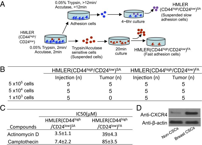
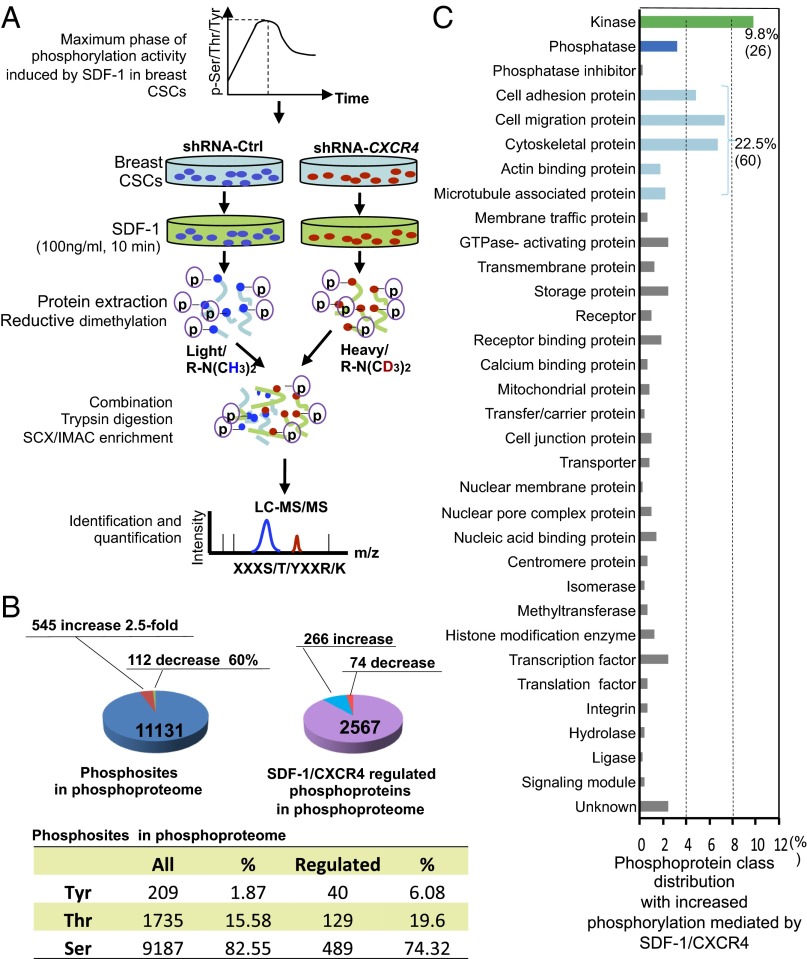
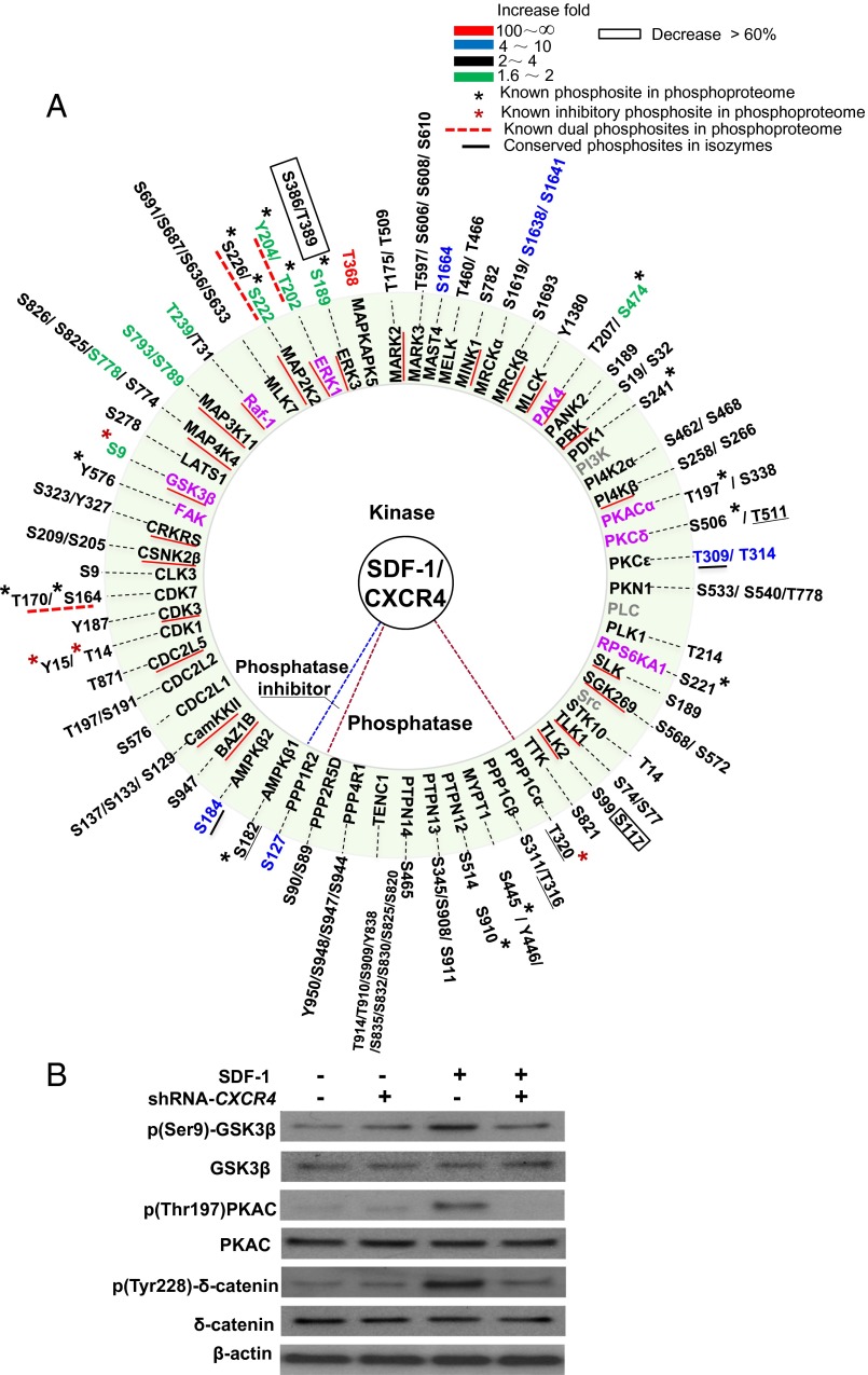
Breast cancer is the leading cause of cancer-related mortality in women worldwide, with an estimated 1.7 million new cases and 522,000 deaths around the world in 2012 alone. Cancer stem cells (CSCs) are essential for tumor reoccurrence and metastasis which is the major source of cancer lethality. G protein-coupled receptor chemokine (C-X-C motif) receptor 4 (CXCR4) is critical for tumor metastasis. However, stromal cell-derived factor 1 (SDF-1)/CXCR4-mediated signaling pathways in breast CSCs are largely unknown. Using isotope reductive dimethylation and large-scale MS-based quantitative phosphoproteome analysis, we examined protein phosphorylation induced by SDF-1/CXCR4 signaling in breast CSCs. We quantified more than 11,000 phosphorylation sites in 2,500 phosphoproteins. Of these phosphosites, 87% were statistically unchanged in abundance in response to SDF-1/CXCR4 stimulation. In contrast, 545 phosphosites in 266 phosphoproteins were significantly increased, whereas 113 phosphosites in 74 phosphoproteins were significantly decreased. SDF-1/CXCR4 increases phosphorylation in 60 cell migration- and invasion-related proteins, of them 43 (>70%) phosphoproteins are unrecognized. In addition, SDF-1/CXCR4 upregulates the phosphorylation of 44 previously uncharacterized kinases, 8 phosphatases, and 1 endogenous phosphatase inhibitor. Using computational approaches, we performed system-based analyses examining SDF-1/CXCR4-mediated phosphoproteome, including construction of kinase-substrate network and feedback regulation loops downstream of SDF-1/CXCR4 signaling in breast CSCs. We identified a previously unidentified SDF-1/CXCR4-PKA-MAP2K2-ERK signaling pathway and demonstrated the feedback regulation on MEK, ERK1/2, δ-catenin, and PPP1Cα in SDF-1/CXCR4 signaling in breast CSCs. This study gives a system-wide view of phosphorylation events downstream of SDF-1/CXCR4 signaling in breast CSCs, providing a resource for the study of CSC-targeted cancer therapy.

Keywords: GPCR; MAPK; chemokine receptor.


Conflict of interest statement

The authors declare no conflict of interest.

Figures

Fig. 1.
Fig. 1.
Enriched breast CSCs expresses high levels of CXCR4. (A) A small percentage of HMLER(CD44high/CD24low)FA cells with trypsin/Accutase sensitive and fast adhesion characters were isolated. (B) HMLER (CD44high/CD24low)FA cells presents potent tumor growth capacity. (C) HMLER (CD44high/CD24low)FA cells show increased drug resistance. (D) Enriched breast CSCs of HMLER (CD44high/CD24low)FA cells express high levels of CXCR4.
Fig. 2.
Fig. 2.
Overview of analyses of SDF-1/CXCR4–induced phosphoproteome in breast CSCs. (A) Schematic of phosphoproteome analyses of the maximum phase of phosphorylation activity induced by SDF-1/CXCR4 signaling in breast CSCs. Breast CSCs with or without transient CXCR4 knockdown [transiently transduced with shRNA-CXCR4 or shRNA-control (Ctrl) plasmids] were treated with 100 ng/mL SDF-1 for 10 min. The purified phosphopeptides were performed isotope reductive dimethylation labeling and enrichment followed by LC-MS/MS analyses. (B) Pie charts show detected phosphosites, phosphoproteins, and SDF-1/CXCR4–regulated phosphoproteins and their distribution. A total of 266 phosphoproteins showed phosphorylation increase ≥2.5-fold. (C) Protein class distribution of 61.4% phosphorylation increased proteins in B. The kinases (26) occupy 9.8% and cell migration-related proteins (60) occupy 22.5%. The other total 38.6% of known proteins with increased phosphorylation and the single-item percentage less than 0.4% are not shown.
Fig. 3.
Fig. 3.
Phosphorylation in kinases, phosphatases, and phosphatase inhibitors. (A) For the proteins, the known kinases in SDF-1/CXCR4 signaling in both are in purple, known kinases in SDF-1/CXCR4 signaling not detected in this phosphoproteome are in gray, and phosphoproteins in phosphoproteome are in black. Recovered kinases (phosphorylation increase 1.6- to ∼2.5-fold) are in black with red underlining. Phosphosites with a black asterisk represent known active phosphosites in phosphoproteome. Phosphosites with a red asterisk represent known inhibitory phosphosites in this phosphoproteome. Known dual phosphosites in phosphoproteome are indicated by red dashed underlining. Conserved phosphosites in isozymes are underlined in black. The color bar in the key indicates phosphorylation increase fold. (B) Western blot analyses of SDF-1/CXCR4 regulated phosphorylation of kinases and effectors in A.
Fig. 4.
Fig. 4.
SDF-1/CXCR4 increases phosphorylation in the established protein complexes. SDF-1/CXCR4 significantly increases phosphorylation at catalytic subunits PPP1C (α/β) and myosin-targeting subunit MYPT1 of MP, catalytic subunit α, anchoring subunit AKAP2/11/12 of PKA, regulatory subunits β1/β2 of energy sensor AMPK, and cell invasion key factor δ-catenin. Black asterisks indicate known active phosphosites in phosphoproteome. Red asterisks indicate known inhibitory phosphosites in this phosphoproteome. Conserved phosphosites in isozymes are underlined in black. Gray circles indicate proteins that are known complex components not in the phosphoproteome.
Fig. 5.
Fig. 5.
An MAPK network downstream of SDF-1/CXCR4 signaling in breast CSCs. The SDF-1/CXCR4–induced MAPK subnetwork shows reconstructed nested pathways with a five-tiered MAPK cascade and known dual phosphosites in MAP2K2 and ERK1. Black arrows show the known phosphorylation relationship in SDF-1/CXCR4 signaling in both. Purple arrows indicate the experimentally confirmed pathway in this study. Blue arrows indicate the known direct interaction and phosphorylation relationship in both. Green arrows represent the indirect phosphorylation relationship in the phosphoproteome. Pink arrows indicate biological function regulation. Red lines indicate dephosphorylation or inhibition. The green circle is the predicted functional complex (specific sites of the components are shown in Fig. 3). The light green MAPK cascade shows the five-tiered MAPK cascade in phosphoproteome.
Fig. 6.
Fig. 6.
Dephosphorylation feedback regulation on MEK1/2, ERK1/2, δ-catenin, and PPP1Cα. (A) Western blot assay of SDF-1 (100 ng/mL) induced phosphorylation turnover of p(Ser217/Ser221)MEK1/2, p(Tyr228)-δ-catenin, and dual phosphosites in p(Thr202/Tyr204)-ERK1/2 over longer time periods in breast CSCs. The relative phosphorylation ratios compared with control are shown. (B) Western blot assays of p(Thr320)-PPP1Cα phosphorylation homeostasis in breast CSCs over time (100 ng/mL SDF-1 treatment). (C) CXCR4 antagonist AMD3100 inhibits SDF-1/CXCR4–induced phosphorylation increase of p(Thr320)-PPP1Cα in breast CSCs. Breast CSCs were pretreated with 30 μg/mL AMD3100 for 1 h followed by with/without 100 ng/mL SDF-1 treatment for 20 min.
Fig. 7.
Fig. 7.
SDF-1/CXCR4-PKA-MAP2K2-ERK pathway in breast CSCs. (A) CXCR4 antagonist AMD3100 inhibits SDF-1/CXCR4–induced phosphorylation increase of p(Thr197)-PKA, p(Ser217/Ser221)MEK1/2, and p(Thr202/Thy204)-ERK1/2 in breast CSCs. (B) PKA inhibitor 14-22-Amide inhibits SDF-1/CXCR4–induced phosphorylation of p(Ser217/Ser221)MEK1/2, and p(Thr202/Thy204)-ERK1/2 in breast CSCs. The breast CSCs cultured in MEBM for overnight were pretreated with 30 μg/mL AMD3100 or 6 µg/mL 14-22-Amide for 1 h followed by with/without SDF-1 (100 ng/mL) treatment for 10 min.

Comment in

References

    1. Al-Hajj M, Wicha MS, Benito-Hernandez A, Morrison SJ, Clarke MF. Prospective identification of tumorigenic breast cancer cells. Proc Natl Acad Sci USA. 2003;100(7):3983–3988. - PMC - PubMed
    1. Müller A, et al. Involvement of chemokine receptors in breast cancer metastasis. Nature. 2001;410(6824):50–56. - PubMed
    1. Tachibana K, et al. The chemokine receptor CXCR4 is essential for vascularization of the gastrointestinal tract. Nature. 1998;393(6685):591–594. - PubMed
    1. Linding R, et al. Systematic discovery of in vivo phosphorylation networks. Cell. 2007;129(7):1415–1426. - PMC - PubMed
    1. Balabanian K, et al. The chemokine SDF-1/CXCL12 binds to and signals through the orphan receptor RDC1 in T lymphocytes. J Biol Chem. 2005;280(42):35760–35766. - PubMed

Publication types

LinkOut - more resources