Review
. 2014 Apr 9:5:141.
doi: 10.3389/fphys.2014.00141. eCollection 2014.

Role of pancreatic stellate cells in chemoresistance in pancreatic cancer

Affiliations
Review

Role of pancreatic stellate cells in chemoresistance in pancreatic cancer

Joshua A McCarroll et al. Front Physiol. .

Abstract

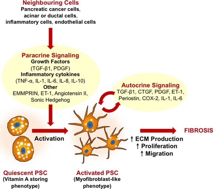
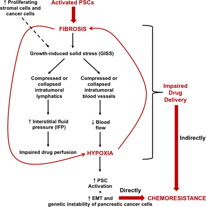
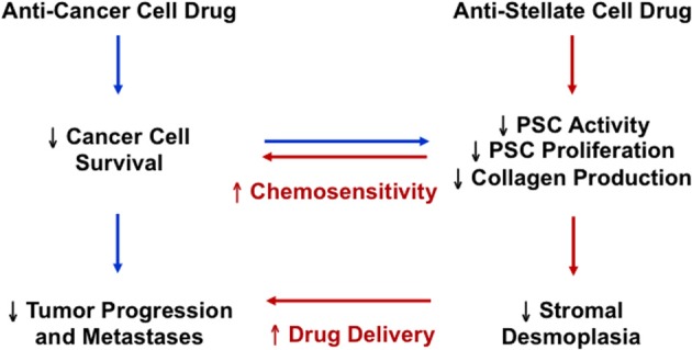
Pancreatic cancer is highly chemoresistant. A major contributing factor is the characteristic extensive stromal or fibrotic reaction, which comprises up to 90% of the tumor volume. Over the last decade there has been intensive research into the role of the pro-fibrogenic pancreatic stellate cells (PSCs) and their interaction with pancreatic cancer cells. As a result of the significant alterations in the tumor microenvironment following activation of PSCs, tumor progression, and chemoresistance is enhanced. This review will discuss how PSCs contribute to chemoresistance in pancreatic cancer.

Keywords: chemoresistance; fibrosis; hypoxia; pancreatic cancer; pancreatic stellate cells; stroma.


Figures

Figure 1
Figure 1
Mechanisms of pancreatic stellate cell (PSC) activation. A central feature of PSCs is the transformation from a quiescent (fat storing phenotype) to an activated (myofibroblast-like phenotype) state. Growth factors and pro-inflammatory cytokines released by neighboring cells (pancreatic cancer cells, injured acinar or ductal cells, inflammatory cells, and endothelial cells) all induce PSC activation. Activated PSCs can then perpetuate this activation state via autocrine stimuli, leading to increased proliferation, migration, and excessive ECM production. In pancreatic cancer, activation of PSCs leads to the production of extensive fibrosis which in turn contributes to disease progression, metastases and chemoresistance. CTGF, connective tissue growth factor; COX-2, cyclooxygenase-2; ECM, extracellular matrix; EMMPRIN, extracellular matrix metalloproteinase inducer; ET-1, endothelin 1; IL, interleukin; PDGF, platelet derived growth factor; TGF-β, transforming growth factor β; TNFα, tumor necrosis factor α; TRAIL, TNF-related apoptosis-inducing ligand.
Figure 2
Figure 2
Growth-induced solid stress (GISS), the hypoxia-fibrosis cycle and their contribution to chemoresistance. Continuous PSC activation results in excessive ECM deposition, particularly of tensile-resistant fibrillar collagen as well as compression-resistant hyaluronan. This eventually leads to prominent fibrosis which, along with deformation caused by the number of proliferating stromal cells and cancer cells, results in GISS. Consequently, this reduces the caliber of intratumoral lymphatics and blood vessels. The former leads to increased IFP, which can impair drug perfusion, while the latter reduces blood flow, consequently leading to intrastromal and intratumoral hypoxia. This loops back onto PSCs, driving their activation and hence generating more fibrosis, which creates a hypoxia–fibrosis cycle. The cycle indirectly contributes to chemoresistance by impairing drug delivery to cancer cells. Moreover, hypoxia is capable of increasing the genetic instability and EMT of pancreatic cancer cells, directly fuelling their chemoresistance. EMT, epithelial-mesenchymal transition.
Figure 3
Figure 3
The potential therapeutic implication of targeting both pancreatic cancer cells and pancreatic stellate cells in pancreatic cancer.

References

    1. Alvarez R., Musteanu M., Garcia-Garcia E., Lopez-Casas P. P., Megias D., Guerra C., et al. (2013). Stromal disrupting effects of nab-paclitaxel in pancreatic cancer. Br. J. Cancer 109, 926–933 10.1038/bjc.2013.415 - DOI - PMC - PubMed
    1. Aoki H., Ohnishi H., Hama K., Ishijima T., Satoh Y., Hanatsuka K., et al. (2006a). Autocrine loop between TGF-beta1 and IL-1beta through Smad3- and ERK-dependent pathways in rat pancreatic stellate cells. Am. J. Physiol. Cell Physiol. 290, C1100–C1108 10.1152/ajpcell.00465.2005 - DOI - PubMed
    1. Aoki H., Ohnishi H., Hama K., Shinozaki S., Kita H., Yamamoto H., et al. (2006b). Existence of autocrine loop between interleukin-6 and transforming growth factor-beta1 in activated rat pancreatic stellate cells. J. Cell. Biochem. 99, 221–228 10.1002/jcb.20906 - DOI - PubMed
    1. Apte M. V., Haber P. S., Applegate T. L., Norton I. D., McCaughan G. W., Korsten M. A., et al. (1998). Periacinar stellate shaped cells in rat pancreas: identification, isolation, and culture. Gut. 43, 128–133 10.1136/gut.43.1.128 - DOI - PMC - PubMed
    1. Apte M. V., Haber P. S., Darby S. J., Rodgers S. C., McCaughan G. W., Korsten M. A., et al. (1999). Pancreatic stellate cells are activated by proinflammatory cytokines: implications for pancreatic fibrogenesis. Gut. 44, 534–541 10.1136/gut.44.4.534 - DOI - PMC - PubMed