Review
. 2014:2014:468309.
doi: 10.1155/2014/468309. Epub 2014 Mar 25.

Optimal management of the critically ill: anaesthesia, monitoring, data capture, and point-of-care technological practices in ovine models of critical care

Affiliations
Review

Optimal management of the critically ill: anaesthesia, monitoring, data capture, and point-of-care technological practices in ovine models of critical care

Saul Chemonges et al. Biomed Res Int. 2014.

Abstract

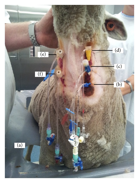
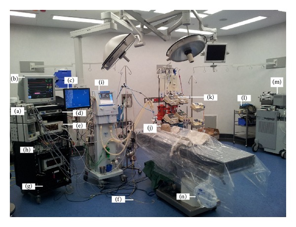
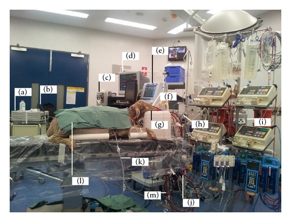
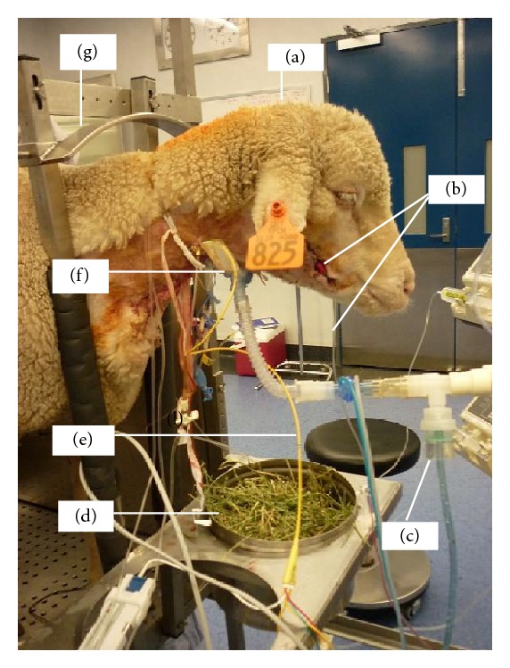
Animal models of critical illness are vital in biomedical research. They provide possibilities for the investigation of pathophysiological processes that may not otherwise be possible in humans. In order to be clinically applicable, the model should simulate the critical care situation realistically, including anaesthesia, monitoring, sampling, utilising appropriate personnel skill mix, and therapeutic interventions. There are limited data documenting the constitution of ideal technologically advanced large animal critical care practices and all the processes of the animal model. In this paper, we describe the procedure of animal preparation, anaesthesia induction and maintenance, physiologic monitoring, data capture, point-of-care technology, and animal aftercare that has been successfully used to study several novel ovine models of critical illness. The relevant investigations are on respiratory failure due to smoke inhalation, transfusion related acute lung injury, endotoxin-induced proteogenomic alterations, haemorrhagic shock, septic shock, brain death, cerebral microcirculation, and artificial heart studies. We have demonstrated the functionality of monitoring practices during anaesthesia required to provide a platform for undertaking systematic investigations in complex ovine models of critical illness.


Figures

Figure 1
Figure 1
An adult Merino ewe being prepared for venovenous extracorporeal membrane oxygenation (VV-ECMO). VV-ECMO is implemented in patients with severe respiratory failure refractory to conventional ventilatory support to provide gas exchange. Venous blood from the patient is accessed from large central veins and returned to the right atrium after it has passed through an oxygenator. The animal has been restrained in a sling cage (a); ventral neck hair has been clipped and aseptically prepared to allow intravenous access to be gained; note the brown colour of povidone iodine. A multilumen central venous catheter has been inserted into the left jugular vein of the animal under local anaesthetic and sutured into place (b) to allow for blood sampling and medications and fluid administration. The left jugular vein has been further cannulated with an 8G sheath for the insertion of a pulmonary artery catheter (c) for haemodynamic monitoring. An 11G sheath catheter has been inserted proximally into the left jugular vein under local anaesthetic and sutured into place to allow for intracardiac echocardiography (ICE) catheter to be inserted (d). The right jugular vein has been cannulated both proximally (e) and distally (f) with single lumen central lines to aid subsequent insertion of return and access ECMO cannulae, respectively. Incremental doses of midazolam are administered to maintain sheep comfort during the prolonged procedure.
Figure 2
Figure 2
Standard animal intensive care unit (ICU) theatre of the Critical Care Research Group (CCRG). For continuous haemodynamic monitoring, the Marquette Solar 8000 Patient ICU CCU Monitoring System (GE Healthcare, Waukesha, WI, USA) consisting of modules (a), displays (b and c), and processor unit (d) together with Vigilance II monitor (e) coupled with Swan-Ganz CCOmbo pulmonary artery catheter (f) (Edwards Lifesciences, Irvine, CA, USA) are used. Other accessories include the hard disc (g) for data storage and an analogue to digital data converter (h). Recruitment of the intensive care ventilator Galileo (Hamilton Medical AG, Switzerland) (i) is performed with the aid of an Ohmeda breathing bag (j) before every experiment. Fluids and medications are delivered from the intravenous fluid workstation (k). Some experiments require the use of an electrosurgical unit (l) and echocardiography (m). All animals have bladder catheters and urine is collected in a sterile bag (n).
Figure 3
Figure 3
Sheep on venovenous extracorporeal membrane oxygenation (VV-ECMO). Equipment includes the Stryker light source (a) attached to an Olympus bronchoscope (b) for bronchoscopy and bronchoalveolar lavage. The Marquette Solar 8000 Patient ICU CCU Monitoring System (GE Healthcare, Waukesha, WI, USA) (c) and monitor (d). The Galileo ICU ventilator (e). ECMO return (f) and access cannulae (b), respectively. The pump assembly consisting of the non-CE marked air-oxygen mixture (FiO2) (Sechrist, Anaheim, CA, USA) (h). CE marked ECMO 550 Bio-Console Bio-Medicus (Medtronic Bio-Medicus Inc., Eden Prairie, MN, USA) (i). CE marked artificial lung (Quadrox PLS) oxygenator (j). Chest drain (K). Sheep (l). Permanent Life Support (PLS) ECMO circuit (MAQUET Cardiopulmonary AG, USA) (m). The Rotaflow Centrifugal Pump and surface coated circuitry, the console pump speed controller (Medtronic) with external drive head, and the heating source are not visible.
Figure 4
Figure 4
Ovine model of smoke inhalation injury. Nebulisation in a conscious sheep (a) in standing position following smoke inhalation via tracheostomy. Anaesthesia was induced using propofol 4 mg/kg IV and maintained with continuous infusion of midazolam 0.7 mg/kg/hr IV and ketamine 8 mg/kg/hr IV to facilitate instrumentation before allowing the sheep to recover. The right facial artery was cannulated to facilitate continuous arterial blood pressure monitoring and serial arterial blood gas analysis (b). Diphoterine or saline was placed in the nebuliser (c) and the sheep had free access to food (d). A Swan-Ganz pulmonary artery catheter (e) was inserted for continuous monitoring of pulmonary artery pressure, central venous pressure, and continuous cardiac output using thermodilution technique. The animal was ventilated through an open surgical tracheostomy (f). The crush (g) prevented the sheep from making large movements that could dislodge the attached instruments. Animals were monitored for up to 21 hours following instrumentation before being sacrificed.
Figure 5
Figure 5
Implanting an artificial heart in an ovine model. This study evaluated a biventricular mechanical device called BiVACOR that is being developed for patients with end stage heart failure. Sheep are anaesthetised and maintained as in humans undergoing cardiac transplantation. In this sheep model, anaesthesia is maintained by constant infusion of propofol and fentanyl. The approach to the heart is via a median sternotomy. Anastomoses to prosthetic blood vessels are created on the pulmonary vein (a) and artery (b) and dorsal aorta and clamped for subsequent attachment to the BiVACOR system (not pictured). In our completed trials, we attached grafts via an end-to-end anastomosis to the ascending aorta (16–18 mm) and pulmonary artery (20 mm) for left and right pump outflows, respectively. Inflow connections were achieved by removing the ventricles just below the atrioventricular groove (leaving about 1 cm of ventricular tissue) and suturing 38 mm grafts (end-to-end) to the ventricular tissue. All grafts were then attached to the pump. Blood flow after the prosthetic heart implant can be monitored by flow meters via probes (c). The surgical exposure of the chest cavity is maintained by a retractor (d) to visualise all organs. The right lung (e), diaphragm, and heart (h) are visible. A stay suture (g) facilitates the suspension of major blood vessels during dissection. Intravenous access for fluid administration was facilitated via a large bore catheter (i). Note the white colour of propofol (j) being used to maintain anaesthesia. During dissection and prior to the removal of the ventricles, continuous haemodynamic monitoring is achieved via the Swan-Ganz pulmonary artery catheter (k).

References

    1. Traber DL, Maybauer MO, Maybauer DM, Westphal M, Traber LD. Inhalational and acute lung injury. Shock. 2005;24(supplement 1):82–87. - PubMed
    1. Shekar K, Fung YL, Diab S, et al. Development of simulated and ovine models of extracorporeal life support to improve understanding of circuit-host interactions. Critical Care and Resuscitation. 2012;14(2):105–111. - PubMed
    1. Hein WR, Griebel PJ. A road less travelled: large animal models in immunological research. Nature Reviews Immunology. 2003;3(1):79–84. - PubMed
    1. Walters EM, Agca Y, Ganjam V, Evans T. Animal models got you puzzled?: think pig. Annals of the New York Academy of Sciences. 2011;1245(1):63–64. - PubMed
    1. Nunoya T, Shibuya K, Saitoh T, et al. Use of miniature pig for biomedical research, with reference to toxicologic studies. Journal of Toxicologic Pathology. 2007;20(3):125–132.

Publication types

LinkOut - more resources